Radiological staging in patients with hilar cholangiocarcinoma: a systematic review and meta-analysis
- PMID: 22919007
- PMCID: PMC3487057
- DOI: 10.1259/bjr/88405305
Radiological staging in patients with hilar cholangiocarcinoma: a systematic review and meta-analysis
Abstract
Objective: To obtain diagnostic performance values of CT, MRI, ultrasound and 18-fludeoxyglucose positron emission tomography (PET)/CT for staging of hilar cholangiocarcinoma.
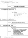
Methods: A comprehensive systematic search was performed for articles published up to March 2011 that fulfilled the inclusion criteria. Study quality was assessed with the quality assessment of diagnostic accuracy studies tool.
Results: 16 articles (448 patients) were included that evaluated CT (n=11), MRI (n=3), ultrasound (n=3), or PET/CT (n=1). Overall, their quality was moderate. The accuracy estimates for evaluation of CT for ductal extent of the tumour was 86%. The sensitivity and specificity estimates of CT were 89% and 92% for evaluation of portal vein involvement, 83% and 93% for hepatic artery involvement, and 61% and 88% for lymph node involvement, respectively. Data were too limited for adequate comparisons of the different techniques.
Conclusion: Diagnostic accuracy studies of CT, MRI, ultrasound or PET/CT for staging of hilar cholangiocarcinoma are sparse and have moderate methodological quality. Data primarily concern CT, which has an acceptable accuracy for assessment of ductal extent, portal vein and hepatic artery involvement, but low sensitivity for nodal status.
Figures
References
-
- Ito F, Cho CS, Rikkers LF, Weber SM. Hilar cholangiocarcinoma: current management. Ann Surg 2009;250:210–18 - PubMed
-
- Cho ES, Park MS, Yu JS, Kim MJ, Kim KW. Biliary ductal involvement of hilar cholangiocarcinoma: multidetector computed tomography versus magnetic resonance cholangiography. J Comput Assist Tomogr 2007;31:72–8 - PubMed
Publication types
MeSH terms
LinkOut - more resources
Full Text Sources
Medical


