. 2012 Nov;153(11):5373-83.
doi: 10.1210/en.2012-1458. Epub 2012 Aug 23.

Estrogen receptors are found in glia and at extranuclear neuronal sites in the dorsal striatum of female rats: evidence for cholinergic but not dopaminergic colocalization

Affiliations

Estrogen receptors are found in glia and at extranuclear neuronal sites in the dorsal striatum of female rats: evidence for cholinergic but not dopaminergic colocalization

Anne Almey et al. Endocrinology. 2012 Nov.

Abstract

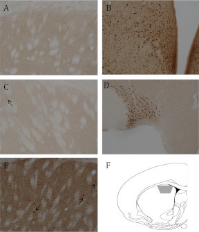
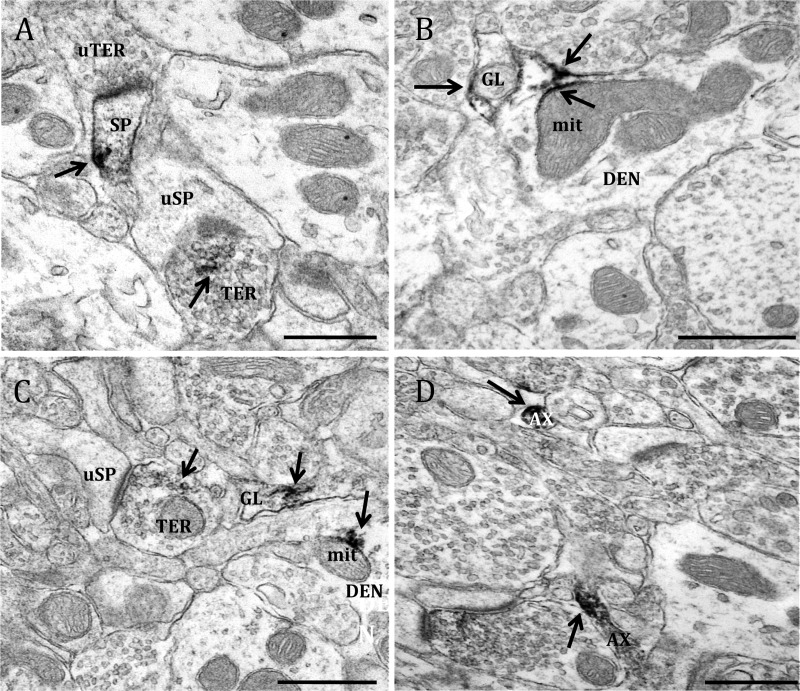
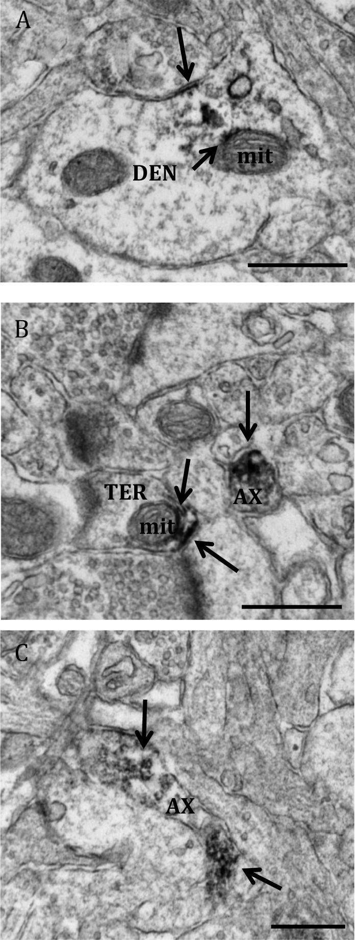
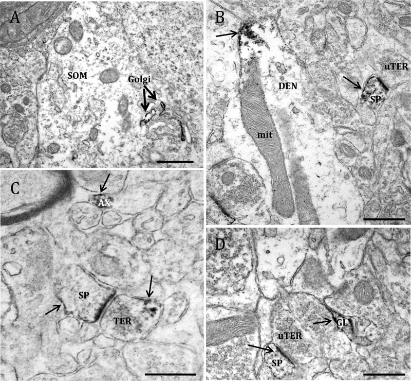
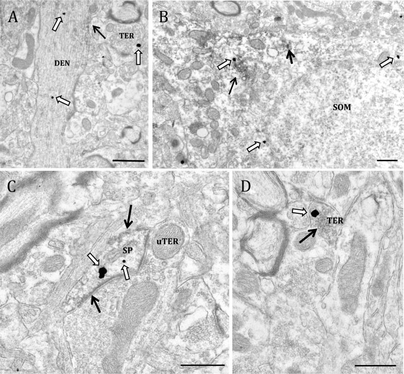
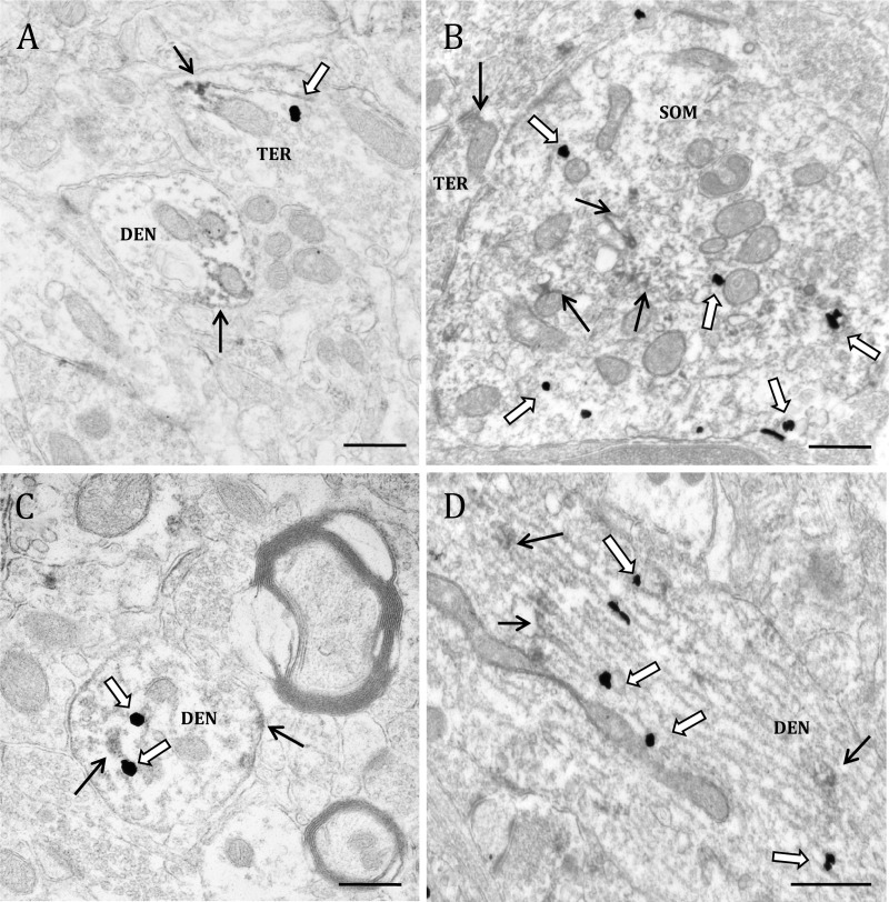
Estrogens rapidly affect dopamine (DA) neurotransmission in the dorsal striatum (dSTR) and DA-related diseases, such as Parkinson's disease and schizophrenia. How estrogens influence DA function remains unclear, in part, because the ultrastructural localization of estrogen receptors (ER) in the dSTR is not known. Light microscopic studies of the dSTR have suggested the presence of ER. This experiment used electron microscopy to determine whether these ER are at extranuclear sites in the dSTR, providing evidence for a mechanism through which estrogen could rapidly affect DA transmission. The dSTR was labeled with antibodies for ERα, ERβ, and G protein-coupled ER 1 (GPER-1) to confirm whether these ER were present in this brain area. After this, the dSTR was dual labeled with antibodies for ERα or GPER-1 and tyrosine hydroxylase or vesicular acetylcholine transporter to determine whether ER are localized to dopaminergic and/or cholinergic processes, respectively. Ultrastructural analysis revealed immunoreactivity (IR) for ERα, ERβ, and GPER-1 exclusively at extranuclear sites throughout the dSTR. ERα-, ERβ-, and GPER-1-IR are mostly frequently observed in axons and glial profiles but are also localized to other neuronal profiles. Dual labeling revealed that ERα- and GPER-1-IR is not associated with DA axons and terminals but is sometimes associated with cholinergic neurons. Because these receptors are exclusively extranuclear in the dSTR, binding at these receptors likely affects neurotransmission via nongenomic mechanisms.


Figures

Fig. 1.
Fig. 1.
Light microscopic localization of ERs. A, Neither nuclear nor extranuclear ERα-IR is detected in the dSTR. B, Dense nuclear ERα-IR in the ventromedial and arcuate nuclei of the hypothalamus. C, No extranuclear ERβ-IR is detected in the dSTR. However, rarely a nucleus with ERβ-IR (arrow) is detected. D, Dense nuclear ERβ-IR in the supraoptic nucleus. E, Dense extranuclear GPER-1-IR is detected in the neuropil of the dSTR. Moreover, several cells with GPER-1-IR (arrows) are seen. F, A coronal schematic of the striatum (atlas level 14; AP 1.00 mm from bregma) (59) indicated the region (gray trapezoid) analyzed.
Fig. 2.
Fig. 2.
Electron micrographs show examples of ERα-containing profiles. ERα-IR is observed in (A) a dendritic spine (SP) that is contacted by an unlabeled axon terminal (uTER), and an axon terminal (TER) that forms an asymmetric synapses with an unlabeled dendritic spine (uSP); (B) a dendritic shaft (DEN), where it is affiliated with the plasma membrane and a mitochondria (mit), and in a glial process (GL); (C) an axon terminal (TER) forming an asymmetric synapse with an unlabeled dendritic spine, a glial profile, and on a mitochondria in a dendritic shaft; and (D) two unmyelinated axons (AX). In this and subsequent figures, labels are placed approximately in the center of the profile, whereas arrows point directly to immunoperoxidase/immunogold labeling. Black arrow, Immunoperoxidase for ERα. Scale bar, 500 μm.
Fig. 3.
Fig. 3.
Electron micrographs show examples of profiles containing ERβ. Rarely, ERβ-IR was detected in (A) a dendritic shaft (DEN) and (B) an axon terminal (TER). Within both profiles, ERβ-IR associated with mitochondria (mit). C, ERβ-IR was observed in unmyelinated axons (AX). Black arrow, Immunoperoxidase for ERβ. Scale bar, 500 μm.
Fig. 4.
Fig. 4.
Electron micrographs showing examples of GPER-1-containing profiles. GPER-1-IR is localized to (A) Golgi bodies (Golgi) in a soma (SOM); (B) a dendritic shaft (DEN) at the plasma membrane, and in a dendritic spine (SP) forming an asymmetric synapse with an unlabeled axon terminal (uTER); (C) an unmyelinated axon (AX) and an axon terminal (TER) forming an asymmetric synapse with a dendritic spine; and (D) a glial process (GL) and dendritic spine contacted by an unlabeled terminal. Black arrow, Immunoperoxidase for GPER-1. Scale bar, 500 μm.
Fig. 5.
Fig. 5.
Electron micrographs show examples of profiles containing ERα and VAChT-IR. A, ERα localized to a VAChT-IR dendrite (DEN) and a VAChT positive terminal (TER). B, A soma (SOM) containing immunogold labeling for VAChT and immunoperoxidase labeling for ERα. C, A dendritic spine (SP) containing ERα-IR and VAChT-IR synapsing onto an unlabeled axon terminal (uTER). D, An axon terminal containing IR for both ERα and VAChT. Black arrow, Immunoperoxidase for ERα; white arrow, immunogold for VAChT. Scale bar, 500 μm.
Fig. 6.
Fig. 6.
Electron micrographs show examples of GPER-1- and VAChT-containing profiles. A, GPER-1 localized to a VAChT-IR terminal (TER) and a dendrite containing GPER-1-IR (DEN). B, A soma (SOM) containing immunogold labeling for VAChT and immunoperoxidase labeling for GPER-1. A terminal containing GPER-1-IR is in apposition to the soma. C, A dendrite that contains GPER-1-IR and VAChT-IR. D, A large dendrite containing IR for both GPER-1 and VAChT. Black arrow, Immunoperoxidase for GPER-1; white arrow, immunogold for VAChT. Scale bar, 500 μm.

Similar articles

Cited by

References

    1. Quinlan MG, Hussain D, Brake WG. 2008. Use of cognitive strategies in rats: the role of estradiol and its interaction with dopamine. Horm Behav 53:185–191 - PubMed
    1. Zurkovsky L, Brown SL, Boyd SE, Fell JA, Korol DL. 2007. Estrogen modulates learning in female rats by acting directly at distinct memory systems. Neuroscience 144:26–37 - PMC - PubMed
    1. Nofrey BS, Ben-Shahar OM, Brake WG. 2008. Estrogen abolishes latent inhibition in ovariectomized female rats. Brain Cogn 66:156–160 - PubMed
    1. Quinlan MG, Duncan A, Loiselle C, Graffe N, Brake WG. 2010. Latent inhibition is affected by phase of estrous cycle in female rats. Brain Cogn 74:244–248 - PubMed
    1. Bourque M, Dluzen DE, Di Paolo T. 2009. Neuroprotective actions of sex steroids in Parkinson's disease. Front Neuroendocrinol 30:142–157 - PubMed

Publication types