

Spectroscopic evaluation of glioma grading at 3T: the combined role of short and long TE
- PMID: 22919334
- PMCID: PMC3417198
- DOI: 10.1100/2012/546171
Spectroscopic evaluation of glioma grading at 3T: the combined role of short and long TE
Abstract
Purpose: To evaluate the diagnostic value of 3T (1)H-MRS in grading cerebral gliomas using short and long echo times.
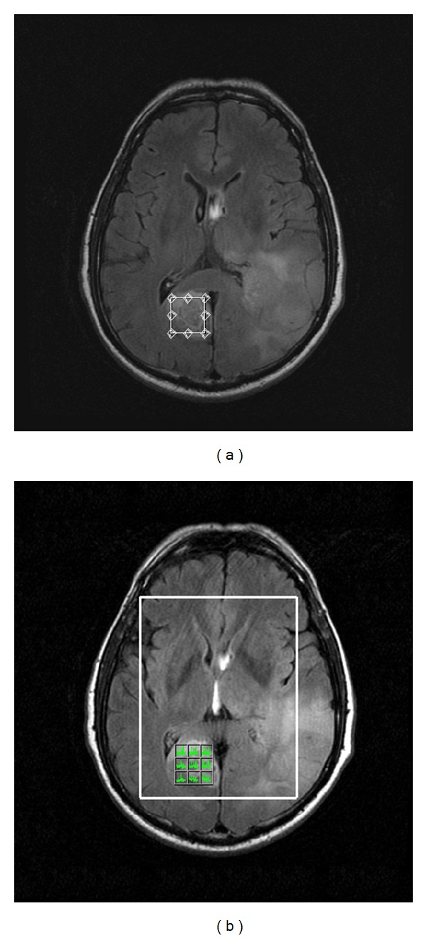
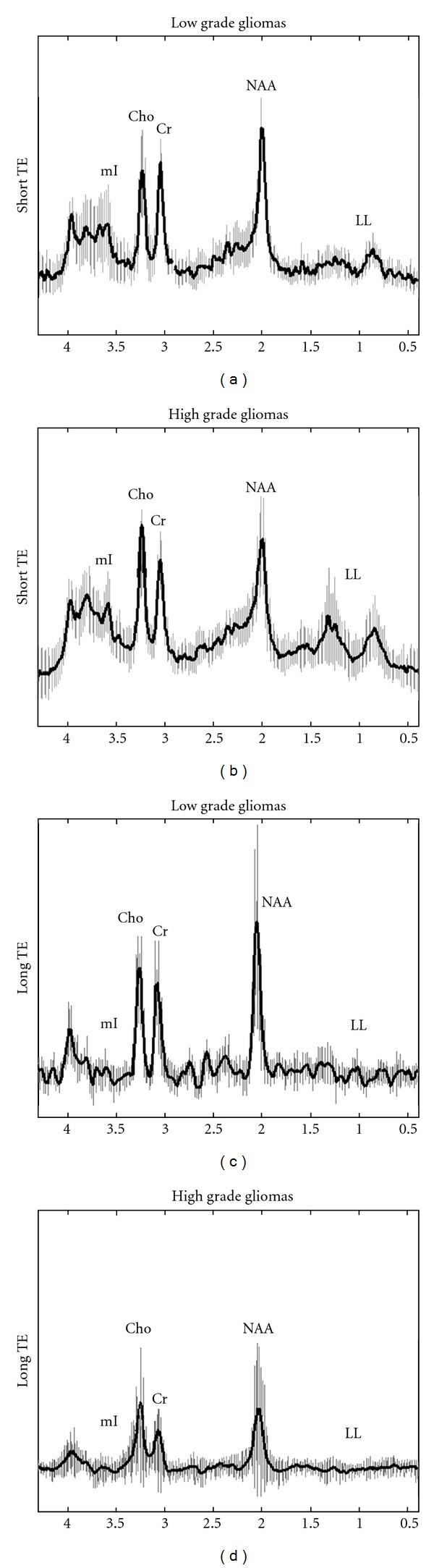
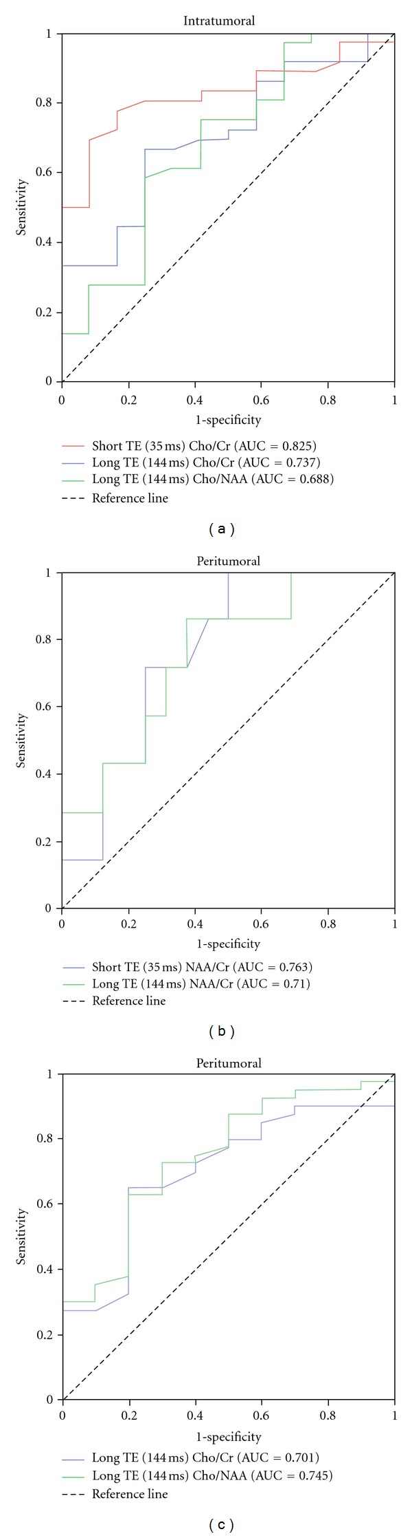
Methods: 1H-MRS was performed on 71 patients with untreated cerebral gliomas. Metabolite ratios of NAA/Cr, Cho/Cr, Cho/NAA, and mI/Cr were calculated for short and long TE and compared between low and high grade gliomas. Lipids were qualitatively evaluated. ROC analysis was performed to obtain the cut-off values for the metabolic ratios presenting statistical difference between the two glioma grades.
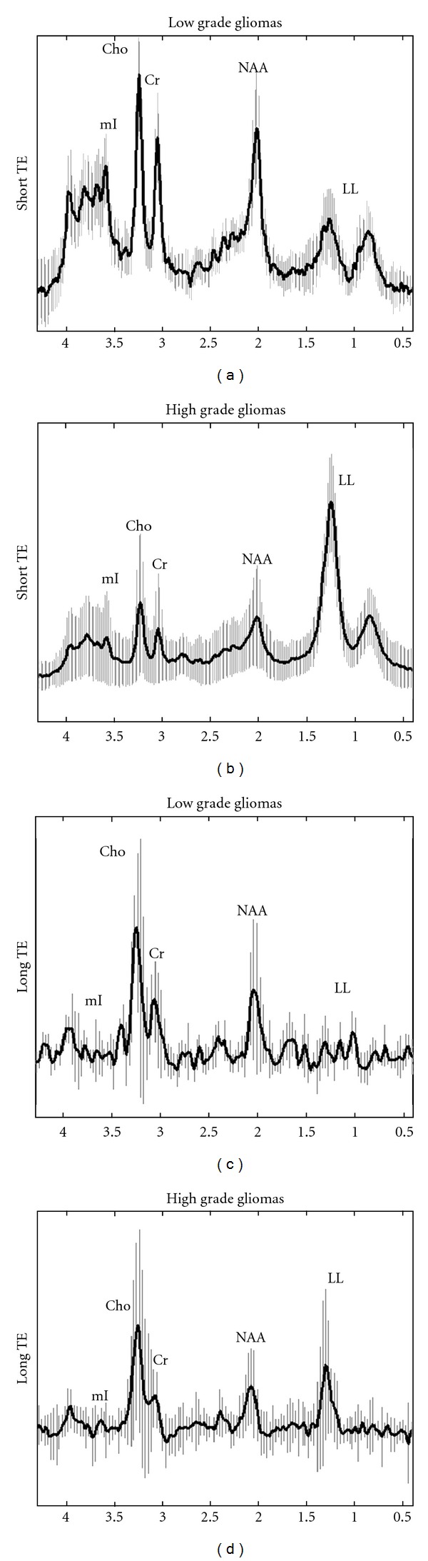
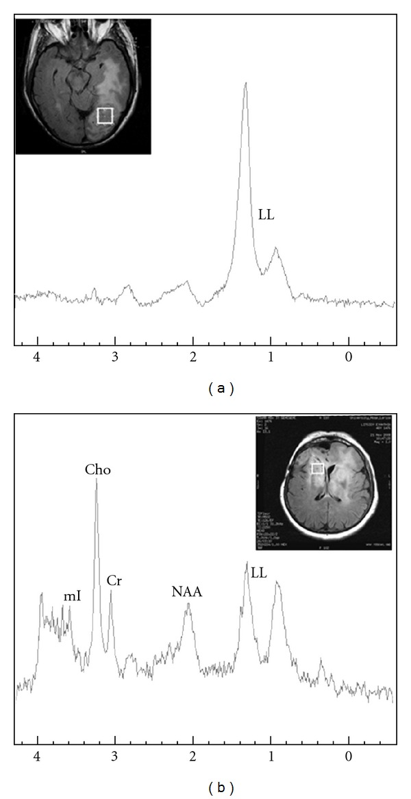
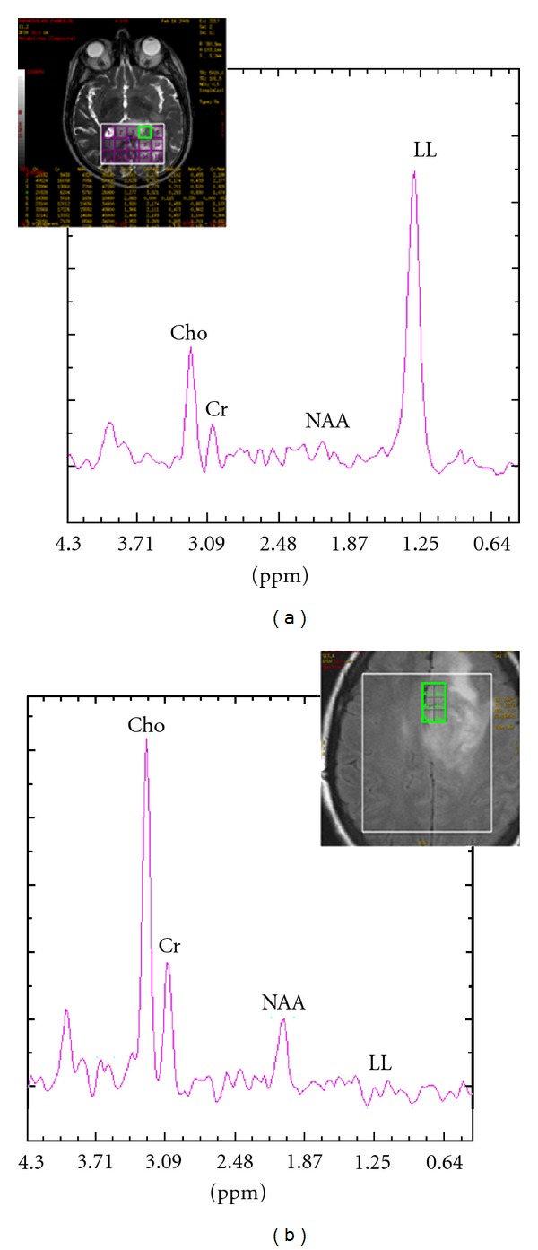
Results: Intratumoral Cho/Cr at both TEs and long TE Cho/NAA were significantly different between low and high grade gliomas. Peritumoral NAA/Cr of both TEs, as well as long TE Cho/Cr and Cho/NAA ratios, significantly differentiated the two tumor grades. Diagnostic sensitivity of peritumoral short TE NAA/Cr proved to be superior over the other metabolic ratios, whereas intratumoral short TE Cho/Cr reached the highest levels of specificity and accuracy. Overall, short TE 1H-MRS reached higher total sensitivity in predicting glioma grade, over long TE.
Conclusion: An advantage was found in using short TE over long TE 1H-MRS in the discrimination of low versus high grade gliomas. Moreover, the results suggested that the peritumoral area of gliomas may be more valuable in predicting glioma grade than using only the intratumoral area.
Figures
References
-
- Martínez-Bisbal MC, Martí-Bonmatí L, Piquer J, et al. 1H and 13C HR-MAS spectroscopy of intact biopsy samples ex vivo and in vivo 1H MRS study of human high grade gliomas. NMR in Biomedicine. 2004;17(4):191–205. - PubMed
-
- Soares DP, Law M. Magnetic resonance spectroscopy of the brain: review of metabolites and clinical applications. Clinical Radiology. 2009;64(1):12–21. - PubMed
-
- Costanzo A, Scarabino T, Trojsi F, et al. Proton MR spectroscopy of cerebral gliomas at 3 T: spatial heterogeneity, and tumour grade and extent. European Radiology. 2008;18(8):1727–1735. - PubMed
-
- Sjøbakk TE, Lundgren S, Kristoffersen A, et al. Clinical 1 H magnetic resonance spectroscopy of brain metastases at 1.5T and 3T. Acta Radiologica. 2006;47(5):501–508. - PubMed
Publication types
MeSH terms
LinkOut - more resources
Full Text Sources
Medical
