

Antioxidant sol-gel improves cutaneous wound healing in streptozotocin-induced diabetic rats
- PMID: 22919368
- PMCID: PMC3420222
- DOI: 10.1155/2012/504693
Antioxidant sol-gel improves cutaneous wound healing in streptozotocin-induced diabetic rats
Abstract
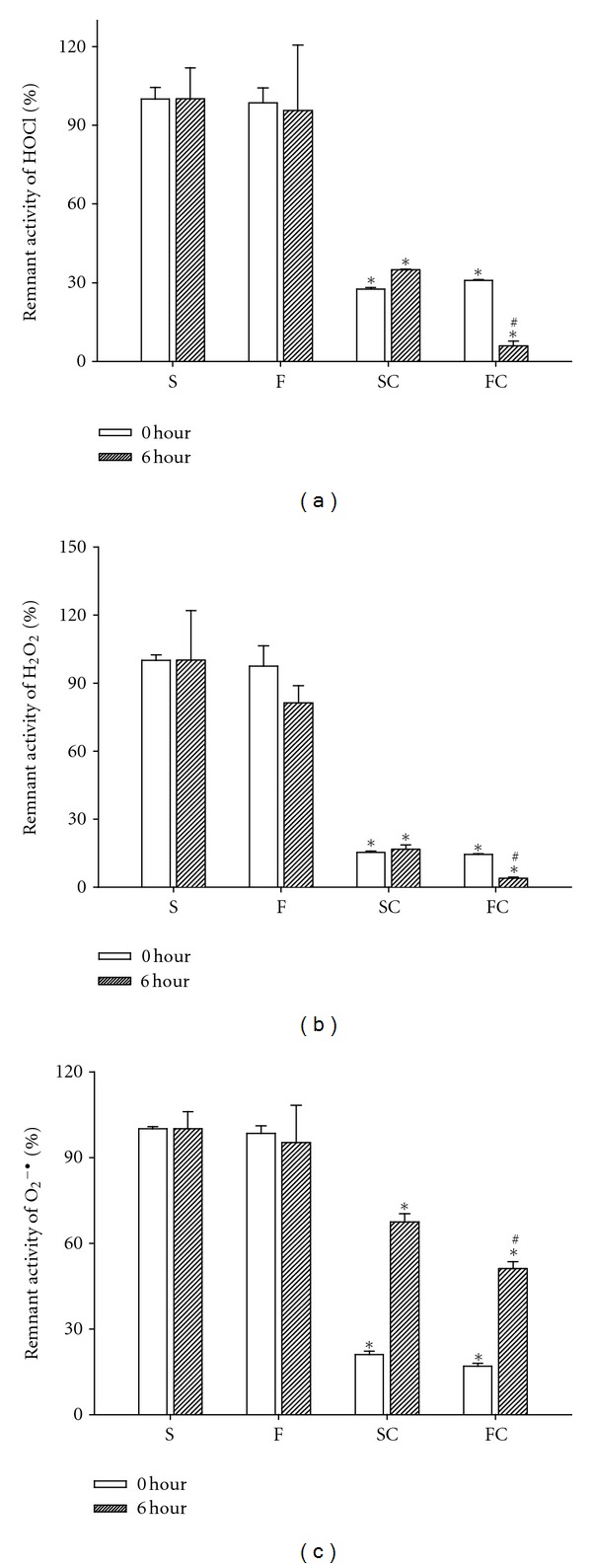
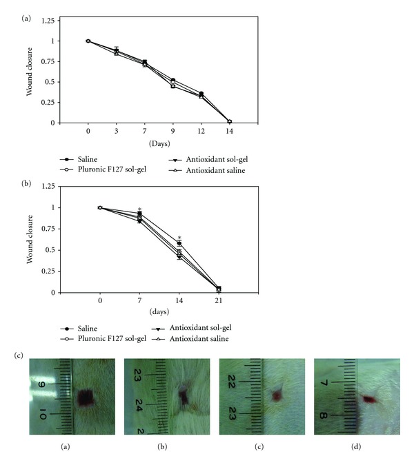
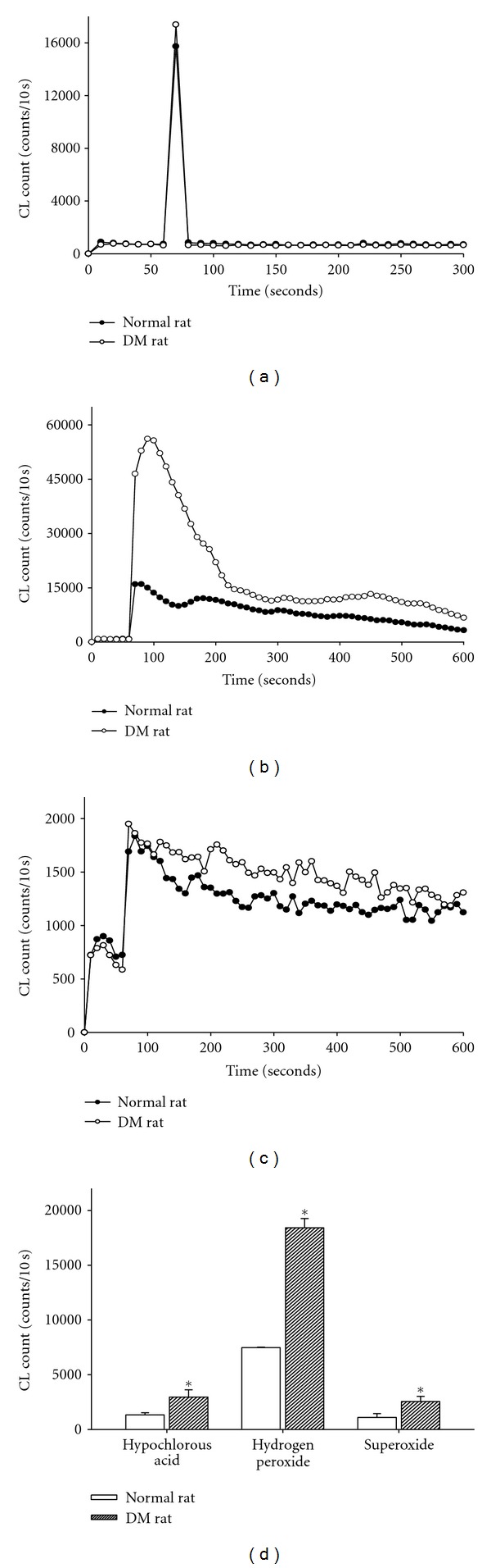
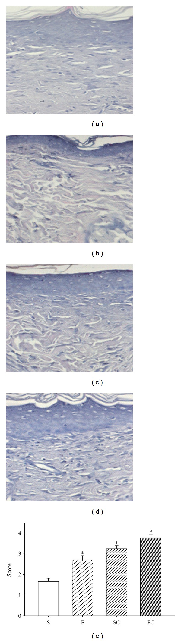
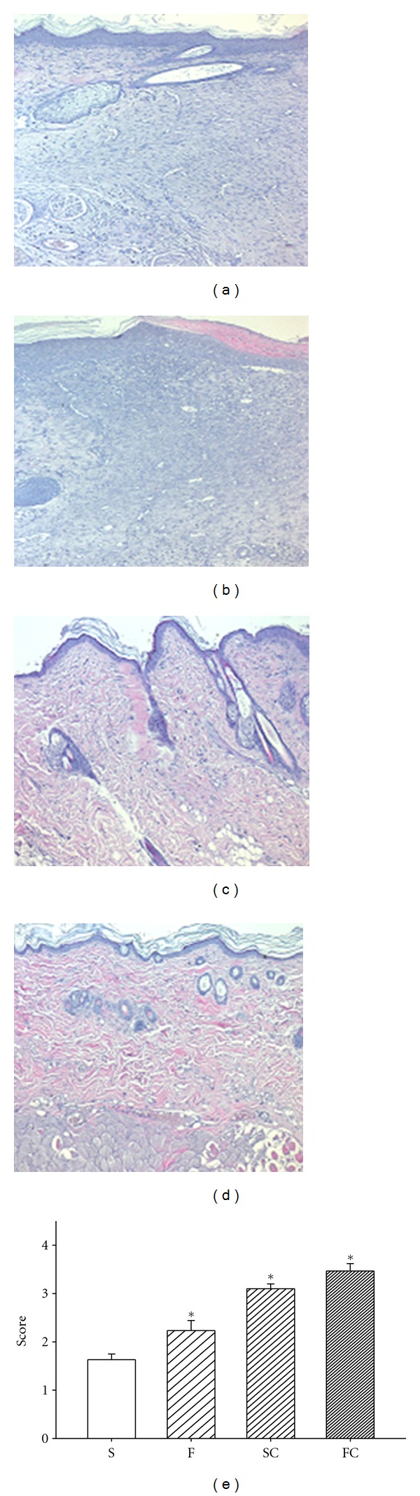
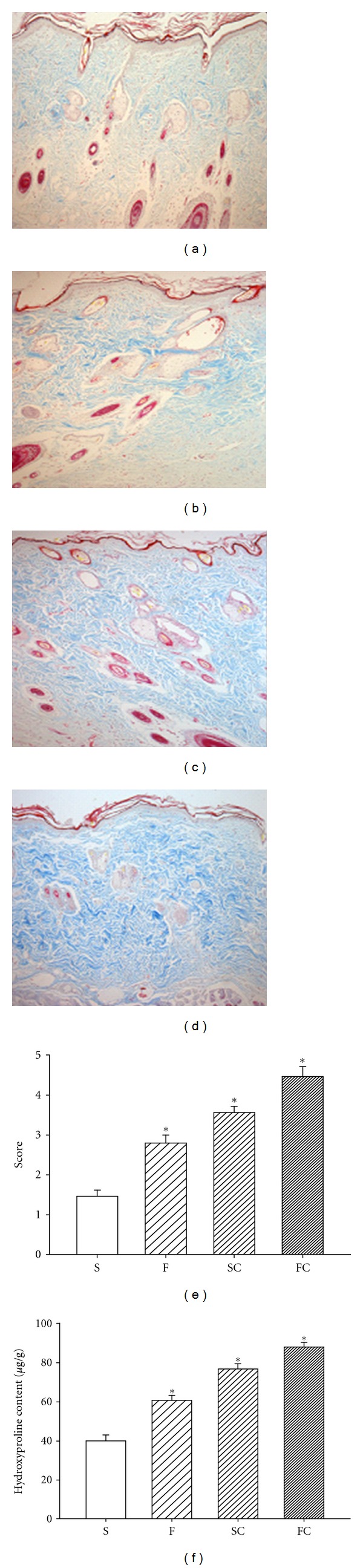
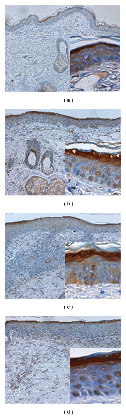
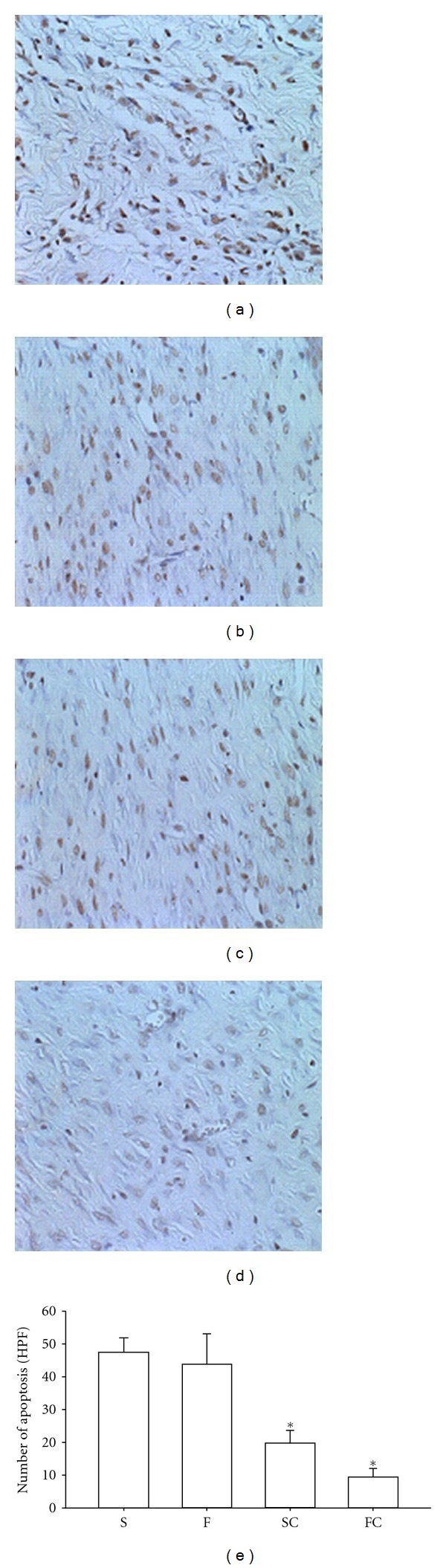
We examined the effects of vitamin C in Pluronic F127 on diabetic wound healing. Full-thickness excision skin wounds were made in normal and diabetic Wistar rats to evaluate the effect of saline, saline plus vitamin C (antioxidant sol), Pluronic F127, or Pluronic F127 plus vitamin C (antioxidant sol-gel). The rate of wound contraction, the levels of epidermal and dermal maturation, collagen synthesis, and apoptosis production in the wound tissue were determined. In vitro data showed that after 6 hours of air exposure, the order of the scavenging abilities for HOCl, H(2)O(2), and O(2) (-) was antioxidant sol-gel > antioxidant saline > Pluronic F127 = saline. After 7 and 14 days of wound injury, the antioxidant sol-gel improved wound healing significantly by accelerated epidermal and dermal maturation, an increase in collagen content, and a decrease in apoptosis formation. However, the wounds of all treatments healed mostly at 3 weeks. Vitamin C in Pluronic F127 hastened cutaneous wound healing by its antioxidant and antiapoptotic mechanisms through a good drug delivery system. This study showed that Pluronic F127 plus vitamin C could potentially be employed as a novel wound-healing enhancer.
Figures
Similar articles
-
Antioxidant and anti-inflammatory potential of curcumin accelerated the cutaneous wound healing in streptozotocin-induced diabetic rats.Int Immunopharmacol. 2014 Jun;20(2):322-30. doi: 10.1016/j.intimp.2014.03.009. Epub 2014 Mar 24. Int Immunopharmacol. 2014. PMID: 24675438
-
Negative air ions through the action of antioxidation, anti-inflammation, anti-apoptosis and angiogenesis ameliorate lipopolysaccharide induced acute lung injury and promote diabetic wound healing in rat.PLoS One. 2022 Oct 26;17(10):e0275748. doi: 10.1371/journal.pone.0275748. eCollection 2022. PLoS One. 2022. PMID: 36288391 Free PMC article.
-
The effect of finger millet feeding on the early responses during the process of wound healing in diabetic rats.Biochim Biophys Acta. 2004 Aug 4;1689(3):190-201. doi: 10.1016/j.bbadis.2004.03.004. Biochim Biophys Acta. 2004. PMID: 15276645
-
Progress in Pluronic F127 Derivatives for Application in Wound Healing and Repair.Int J Nanomedicine. 2023 Aug 7;18:4485-4505. doi: 10.2147/IJN.S418534. eCollection 2023. Int J Nanomedicine. 2023. PMID: 37576462 Free PMC article. Review.
-
[Vitamin C and its derivatives in maintaining the good skin condition].Postepy Biochem. 2024 Sep 17;70(3):307-314. doi: 10.18388/pb.2021_554. Print 2024 Sep 30. Postepy Biochem. 2024. PMID: 39365570 Review. Polish.
Cited by
-
Preventive effects of royal jelly against anaphylactic response in a murine model of cow's milk allergy.Pharm Biol. 2017 Dec;55(1):2145-2152. doi: 10.1080/13880209.2017.1383487. Pharm Biol. 2017. PMID: 28982287 Free PMC article.
-
Sirtuin-6 deficiency exacerbates diabetes-induced impairment of wound healing.Exp Dermatol. 2015 Oct;24(10):773-8. doi: 10.1111/exd.12762. Epub 2015 Aug 18. Exp Dermatol. 2015. PMID: 26010430 Free PMC article.
-
The effects of caffeine on wound healing.Int Wound J. 2016 Oct;13(5):605-13. doi: 10.1111/iwj.12327. Epub 2014 Jul 8. Int Wound J. 2016. PMID: 25041108 Free PMC article.
-
New Amorphous Hydrogels with Proliferative Properties as Potential Tools in Wound Healing.Gels. 2022 Sep 21;8(10):604. doi: 10.3390/gels8100604. Gels. 2022. PMID: 36286105 Free PMC article.
-
Boron promotes streptozotocin-induced diabetic wound healing: roles in cell proliferation and migration, growth factor expression, and inflammation.Mol Cell Biochem. 2016 Jun;417(1-2):119-33. doi: 10.1007/s11010-016-2719-9. Epub 2016 May 20. Mol Cell Biochem. 2016. PMID: 27206737
References
-
- Singer AJ, Clark RAF. Cutaneous wound healing. The New England Journal of Medicine. 1999;341(10):738–746. - PubMed
-
- Luo JD, Wang YY, Fu WL, Wu J, Chen AF. Gene therapy of endothelial nitric oxide synthase and manganese superoxide dismutase restores delayed wound healing in type 1 diabetic mice. Circulation. 2004;110(16):2484–2493. - PubMed
-
- Schorah CJ, Downing C, Piripitsi A, et al. Total vitamin C, ascorbic acid, and dehydroascorbic acid concentrations in plasma of critically ill patients. American Journal of Clinical Nutrition. 1996;63(5):760–765. - PubMed
-
- Clark RAF. Oxidative stress and “senescent” fibroblasts in non-healing wounds as potential therapeutic targets. Journal of Investigative Dermatology. 2008;128(10):2361–2364. - PubMed
-
- Schneir M, Ramamurthy N, Golub L. Dietary ascorbic acid normalizes diabetes-induced underhydroxylation of nascent type I collagen molecules. Collagen and Related Research. 1985;5(5):415–422. - PubMed
Publication types
MeSH terms
Substances
LinkOut - more resources
Full Text Sources
Medical