doi: 10.4103/1995-705X.99227.
Giant left atrium: a review
Affiliations
- PMID: 22919448
- PMCID: PMC3424779
- DOI: 10.4103/1995-705X.99227
Item in Clipboard
Giant left atrium: a review
Heart Views.
2012 Apr.
Abstract
Giant left atrium is a rare condition, with a reported incidence of 0.3%, and following mainly rheumatic mitral valve disease. Although rheumatic heart disease represents the main cause of giant left atrium, other etiologies have been reported. Giant left atrium has significant hemodynamic effects and requires specific management. In this review, we present two cases, discuss the different definitions, etiologies, clinical presentation and management modalities.
Keywords: Atrial fibrillation; atrial plication; compression; giant left atrium; maze procedure; rheumatic heart disease.
Conflict of interest statement
Figures
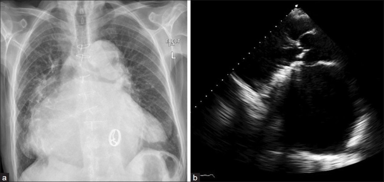
(a) The chest X-ray AP view showed increased cardiothoracic ratio; the left atrial shadow extended towards the left and right cardiac borders, with bilateral lung haziness. The mitral prothesis is visualized. (b) Transthoracic echocardiography in parasternal long axis view showed the proshtetic mitral valve and a markedly dilated left atrium measuring 10.8 cm
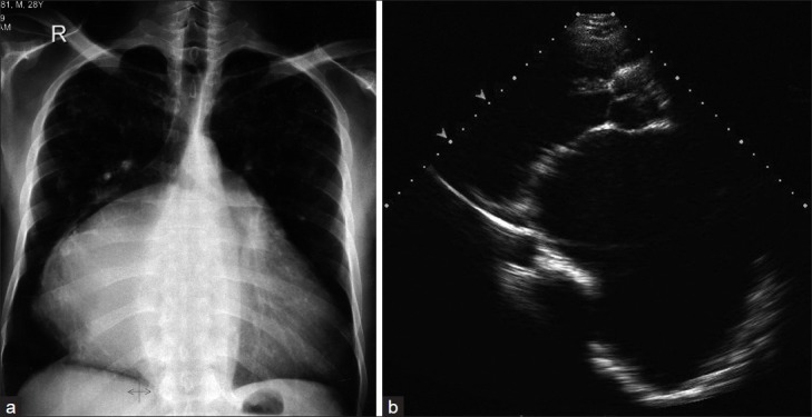
Pre-operative images. (a) The chest X-ray AP view showed increased cardiothoracic ratio, the left atrial shadow is shifted towards the right sternal border with splaying and lifting of the carina as well as the right and left bronchus. (b) Transthoracic echocardiography in parasternal long axis view, revealed a markedly dilated left atrium measuring 12.1 cm with severe mitral stenosis
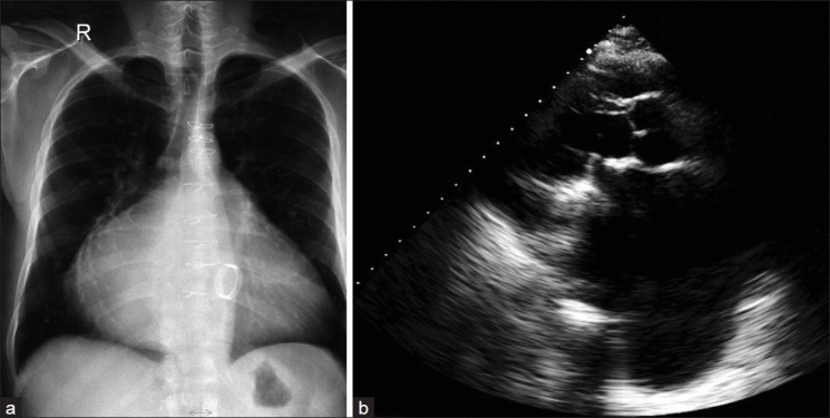
Post-operative images. (a) the chest X-ray AP view and (b) TTE showed mitral prothesis. The dilated left atrium decreased to 10 cm by TTE measurement
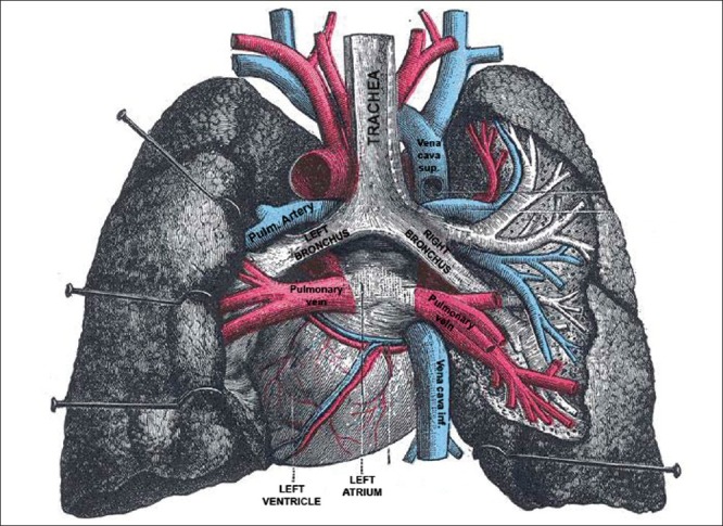
Anatomical specimen showing the left atrium seen from the posterior aspect and the relations to the main, left and right bronchus, the pulmonary veins, systemic veins and both lungs after removal of the esophagus. (Extracted from Gray's Anatomy 20th edition)
References
-
- Wang Y, Gutman JM, Heilbron D, Wahr D, Schiller NB. Atrial volume in a normal adult population by two-dimensional echocardiography. Chest. 1984;86:595–601. - PubMed
-
- Hurst W. Memories of patients with a giant left atrium. Circulation. 2001;104:2630–1. - PubMed
-
- Piccoli GP, Massini C, Di Eusanio G, Ballerini L, Tacobone G, Soro A, et al. Giant left atrium and mitral valve disease: Early and late results of surgical treatment in 40 cases. J Cardiovasc Surg. 1984;25:328–36. - PubMed
-
- Oh JK. Echocardiographic evaluation of morphological and hemodynamic significance of giant left atrium. Circulation. 1992;86:328–30. - PubMed
-
- Owen I, Fenton WJ. A case of extreme dilatation of the left auricle of the heart. Trans Clin Soc London. 1901;34:183–91.