

doi: 10.4302/plp.2011.4.02.
Imaging limbal and scleral vasculature using Swept Source Optical Coherence Tomography
Affiliations
- PMID: 22919461
- PMCID: PMC3423982
- DOI: 10.4302/plp.2011.4.02
Item in Clipboard
Imaging limbal and scleral vasculature using Swept Source Optical Coherence Tomography
Photonics Lett Pol.
.
Abstract
We demonstrate an application of high-speed swept source optical coherence tomography for vessel visualization in the anterior segment of the human eye. The human corneo-scleral junction and sclera was imaged in vivo. Imaging was performed using a swept source OCT system operating at the 1050nm wavelength range and 100kHz A-scan rate. High imaging speed enables the generation of 3D depth-resolved vasculature maps. The vessel visualization method revealed a rich vascular system in the conjunctiva and episclera.
Figures
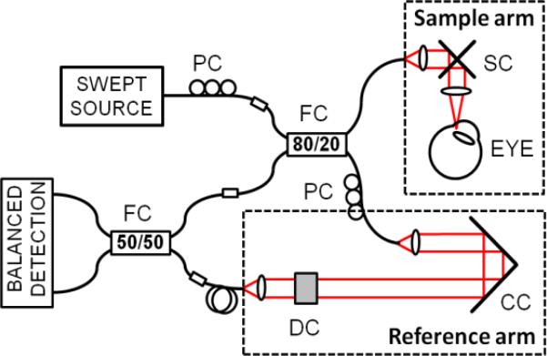
Swept source OCT system. PC – polarization controller, FC – fiber coupler, SC – galvanometric scanners, CC – corner cube, DC – dispersion compensation.
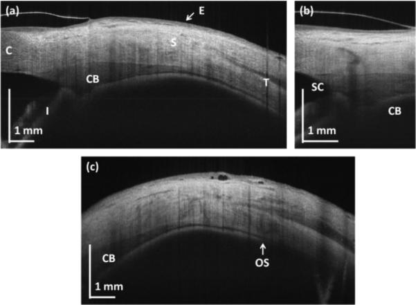
OCT cross-sections of the human corneo-scleral region. (a) Structural high-definition scan (protocol A); (b) limbus (protocol B); (c) peripheral sclera (protocol A). C – cornea, S – sclera, E – epithelium, I – iris, SC – Schlemm's canal, OS – ora serrata.
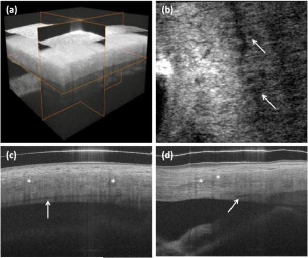
In vivo 3-D imaging of human limbus. (a) rendering and 3-D reconstruction of data set. (b) C-scan (x-y cross section). Arrows show Schlemm's canal. (c) B-scan in fast scanning axis (y-z cross-section). (d) B-scan in slow scanning axis (x-z cross section). Protocol C from Table 1 was used for scanning the eye. Asterisks show blood vessel shadows.
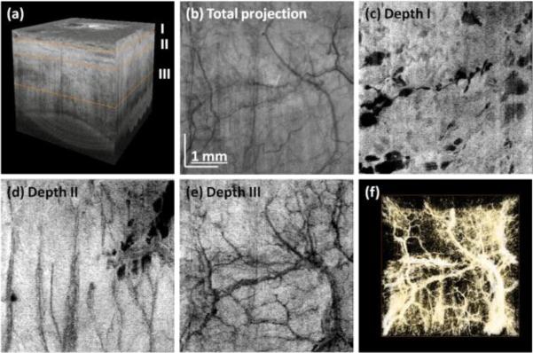
Three-dimensional reconstruction of human limbus after flattening (a). En face projection of structural images (b). Virtual C-scans (projections) from angiographic data set showing vascular networks in episclera (c) and deeper sclera (d)-(e). Vasculature rendering after thresholding and colorscale inversion (f). Data set was acquired using protocol D.
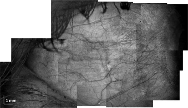
Large area scleral OCT imaging. Composite projection image from 12 individual data sets acquired using protocol D.
References
-
- Drexler W, Fujimoto JG, editors. Optical Coherence Tomography. Technology and Applications. Springer; Berlin-Heidelberg: 2008.
-
- Zysk AM, Oldenburg AL, Marks DL, Nguyen FT, Boppart SA. J. Biomed. Opt. 2007;12:051403. - PubMed
Grants and funding
LinkOut - more resources
Full Text Sources
Other Literature Sources