. 2012:2012:283974.
doi: 10.1155/2012/283974. Epub 2012 Aug 2.

ASGR1 and ASGR2, the Genes that Encode the Asialoglycoprotein Receptor (Ashwell Receptor), Are Expressed in Peripheral Blood Monocytes and Show Interindividual Differences in Transcript Profile

Affiliations

ASGR1 and ASGR2, the Genes that Encode the Asialoglycoprotein Receptor (Ashwell Receptor), Are Expressed in Peripheral Blood Monocytes and Show Interindividual Differences in Transcript Profile

Rebecca Louise Harris et al. Mol Biol Int. 2012.

Abstract

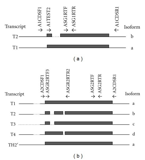
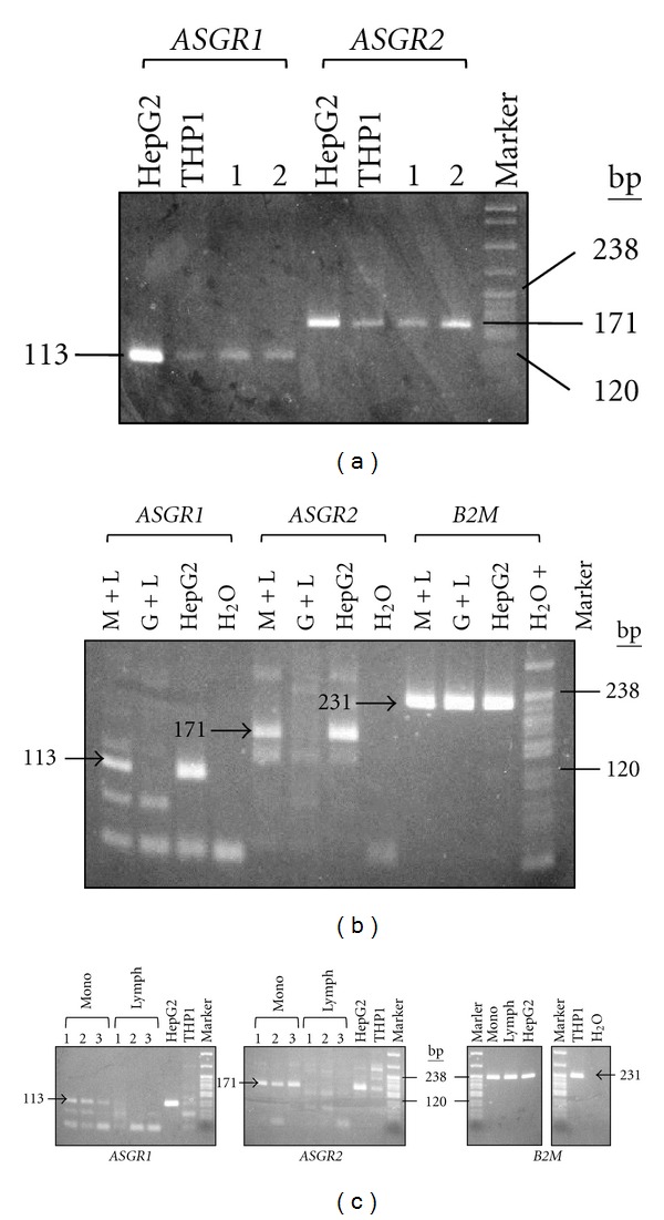
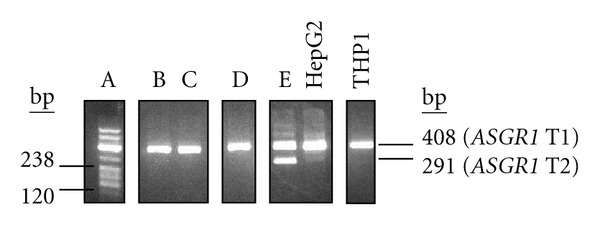
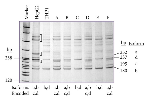
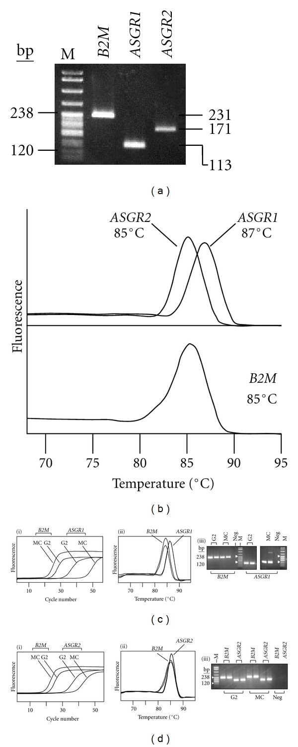
Background. The asialoglycoprotein receptor (ASGPR) is a hepatic receptor that mediates removal of potentially hazardous glycoconjugates from blood in health and disease. The receptor comprises two proteins, asialoglycoprotein receptor 1 and 2 (ASGR1 and ASGR2), encoded by the genes ASGR1 and ASGR2. Design and Methods. Using reverse transcription amplification (RT-PCR), expression of ASGR1 and ASGR2 was investigated in human peripheral blood monocytes. Results. Monocytes were found to express ASGR1 and ASGR2 transcripts. Correctly spliced transcript variants encoding different isoforms of ASGR1 and ASGR2 were present in monocytes. The profile of transcript variants from both ASGR1 and ASGR2 differed among individuals. Transcript expression levels were compared with the hepatocyte cell line HepG2 which produces high levels of ASGPR. Monocyte transcripts were 4 to 6 orders of magnitude less than in HepG2 but nonetheless readily detectable using standard RT-PCR. The monocyte cell line THP1 gave similar results to monocytes harvested from peripheral blood, indicating it may provide a suitable model system for studying ASGPR function in this cell type. Conclusions. Monocytes transcribe and correctly process transcripts encoding the constituent proteins of the ASGPR. Monocytes may therefore represent a mobile pool of the receptor, capable of reaching sites remote from the liver.


Figures

Figure 1
Figure 1
ASGR1 and ASGR2 transcripts and encoded protein isoforms. (a) ASGR1. (b) ASGR2. In both panels solid lines indicate nucleotide sequence that is present in a transcript, grey boxes represent coding sequence, dotted lines indicate sequence that is not present in a transcript. Arrows indicate location and direction of primers used in RT-PCR. Data for transcripts and isoforms are taken from NCBI [14, 15].
Figure 2
Figure 2
Detection of ASGR1 and ASGR2 transcripts in peripheral blood mononuclear cells and localization to monocytes. Transcripts were detected using RT-PCR, the presence of ASGR1 transcripts was indicated by a 113 bp amplification product (primers ASG1RTF + ASG1RTR) and ASGR2 transcripts by a 171 bp product (primers ASG2RTF + ASG2RTR). HepG2 was used as a positive control and water as a negative control. Where necessary, β-2-microglobulin (B2M) was used as a reference to demonstrate equivalent amplification for each RNA preparation, (primers B2MRTF + B2MRTR, product 231 bp). (a) Analysis of RNA from monocyte cell line THP1 and PBMCs from two unrelated individuals (1 and 2). ASGR1 and ASGR2 transcripts were detected in all cases. (b) Analysis of RNA from peripheral blood cell fractions: monocytes + lymphocytes (M + L) and granulocytes + lymphocytes (G + L). ASGR1 and ASGR2 transcripts were detected in M + L but not in G + L. (c) Analysis of RNA from cell-sorted monocytes (mono, lanes 1–3 triplicate analyses) and lymphocytes (lymph, lanes 1–3 triplicate analyses) (data for water control not shown for ASGR1 and ASGR2). The data were obtained from a single experiment involving several gels: these are indicated by separate gel windows. For each panel, analysis was done using agarose gel electrophoresis with pBR322/MspI size standard (“Marker”, 238 bp and 120 bp indicated).
Figure 3
Figure 3
ASGR1 transcript profiles in different individuals. Nested PCR was used to detect ASGR1 transcripts T1 and T2 (first round primers A1CDSF1 + A1CDSR1; second round A1TEST2 + ASG1RTR). A product of 408 bp represented T1, present in RNA from the M + L cell fraction of five individuals (A–E) and also in HepG2 and THP1; a 291 bp product indicated the presence of T2 (individual E, and, very faintly, in HepG2). The RT-PCR product for individual A was coloaded with the DNA size standard (pBR322/MspI, 238 bp and 120 bp sizes indicated). Analysis was done using agarose gel electrophoresis. The figure is a composite of various data (indicated by individual gel windows).
Figure 4
Figure 4
ASGR2 transcript profiles in different individuals. Primer pair ASGR2RTF3 + ASGR2RTR2 gave RT-PCR products of different length (arrowed) according to the ASGR2 transcripts present. RNA extracted from the M + L cell fraction of 6 different individuals (A–F) gave either a profile consistent with transcripts encoding all isoforms (a, b, c, and d) or just with isoforms b and d. The liver cell line HepG2 showed all transcripts, the monocyte cell line THP1 showed only transcripts encoding isoforms b and d. The analysis was done using polyacrylamide gel electrophoresis. DNA size reference was pBR322/MspI (“Marker”, 238 bp and 120 bp sizes indicated). In addition to the expected products, additional major bands were visualized on polyacrylamide gel electrophoresis of the RT-PCR screen for ASGR2 transcripts (bracketed bands). It was considered possible that these represented heteroduplexes arising by cross-hybridization of complementary strands of the closely homologous true PCR products. To test this, each candidate heteroduplex band was excised from the gel, eluted into water, reamplified using the primers used in the initial PCR and the products electrophoresed on polyacrylamide. The results confirmed that the additional bands were hybrid duplexes containing complementary strands from nonidentical true PCR products: each hybrid yielded products consistent with amplification of two different template strands (data not shown).
Figure 5
Figure 5
Real-time PCR analyses. (a) Agarose gel electrophoresis of products from optimized real-time PCRs for ASGR1 (113 bp, primers ASG1RTF + ASG1RTR), ASGR2 (171 bp, primers ASG2RTF + ASG2RTR), and B2M (231 bp, primers B2MRTF + B2MRTR). Substrate was HepG2 cDNA. (b) Melting curve analysis of real-time PCR products from panel (a). The Tm for each product is indicated. (c and d) Relative quantification of ASGR1 (c) and ASGR2 (d) transcripts in cell-sorted monocytes (MC) compared with HepG2 (G2). B2M was used as the reference. (i) Fluorescence profiles obtained during real-time PCR. Samples were analyzed in duplicate, for clarity only one of each duplicate is shown. (ii) Melting curve analysis for the real-time PCR products in (i). (iii) Agarose gel electrophoresis of real-time PCR products (duplicate analyses shown). Neg indicates real-time PCR control containing water instead of nucleic acid template. In panels a, c(iii), and d(iii), M denotes pBR322/MspI size standard (238 bp and 120 bp sizes indicated).

Similar articles

Cited by

References

    1. Weigel PH, Yik JHN. Glycans as endocytosis signals: the cases of the asialoglycoprotein and hyaluronan/chondroitin sulfate receptors. Biochimica et Biophysica Acta. 2002;1572(2-3):341–363. - PubMed
    1. Ellies LG, Ditto D, Levy GG, et al. Sialyltransferase ST3Gal-IV operates as a dominant modifier of hemostasis by concealing asialoglycoprotein receptor ligands. Proceedings of the National Academy of Sciences of the United States of America. 2002;99(15):10042–10047. - PMC - PubMed
    1. Grewal PK, Uchiyama S, Ditto D, et al. The Ashwell receptor mitigates the lethal coagulopathy of sepsis. Nature Medicine. 2008;14(6):648–655. - PMC - PubMed
    1. Zijderhand-Bleekemolen JE, Schwartz AL, Slot JW, Strous GJ, Geuze HJ. Ligand- and weak base-induced redistribution of asialoglycoprotein receptors in hepatoma cells. Journal of Cell Biology. 1987;104(6):1647–1654. - PMC - PubMed
    1. Ii M, Kurata H, Itoh N, Yamashina I, Kawasaki T. Molecular cloning and sequence analysis of cDNA encoding the macrophage lectin specific for galactose and N-acetylgalactosamine. The Journal of Biological Chemistry. 1990;265(19):11295–11298. - PubMed

LinkOut - more resources