. 2012:2:39.
doi: 10.4103/2156-7514.99151. Epub 2012 Jul 28.

Musculoskeletal disorders in the elderly

Affiliations

Musculoskeletal disorders in the elderly

Ramon Gheno et al. J Clin Imaging Sci. 2012.

Abstract

Musculoskeletal disorders are among the most common problems affecting the elderly. The resulting loss of mobility and physical independence can be particularly devastating in this population. The aim of this article is to present some of the most frequent musculoskeletal disorders of the elderly, such as fractures, osteoporosis, osteoarthritis, microcrystal disorders, infections, and tumors.

Keywords: Aged; arthritis; fracture; musculoskeletal diseases; neoplasm.


Conflict of interest statement

Conflict of Interest: None declared.

Figures

Figure 1
Figure 1
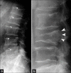
Usual benign vertebral compression (a) multiple levels, diffuse radiolucency, and whole vertebral endplate involvement (arrows); usual malignant vertebral compression characteristics (b) single vertebral involvement, focal and bulging of the posterior wall (arrowheads).
Figure 2
Figure 2
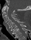
Missed fracture of the odontoid process and vertebral body of C2 (arrows) in a patient with a history of fall one month before. Note the associated degenerative changes of the lower cervical spine (arrowheads).
Figure 3
Figure 3
Transverse fracture of thoracic vertebral body (arrow) and posterior arch (arrowhead) with posterior epidural hematoma (curved arrow) in an ankylosed spine depicted on (a) T1- and (b) T2-weighted sequences.
Figure 4
Figure 4
Insufficiency fractures (arrows) located in the sacrum with adjacent edema (arrowheads), better depicted on (a) T1- and (b) T2- weighted sequences, respectively. This bone edema should not be confused with sacroiliitis.
Figure 5
Figure 5
Multiple fractures (white arrows) in (a) the proximal femur and (b) pubic ramus on coronal T1-weighted images
Figure 6
Figure 6
Subchondral fracture of the femoral head: (a) conventional radiography and (b) fat saturated sagittal T2-weighted image (white arrow).
Figure 7
Figure 7
Typical osteoarthritis of (a) the distal and proximal interphalangeal joints and (b) hip joint: joint space narrowing (arrows), subchondral cysts (arrowheads), osteophytes (curved arrows), and subchondral sclerosis (thick arrow).
Figure 8
Figure 8
Ultrasound images in (a) the long and (b) short axis of the supraspinatus tendon demonstrating a complete tear (arrowheads).
Figure 9
Figure 9
Gout of the first MTP joint in conventional radiography: Excentric dense nodular soft tissue mass (arrows), large erosions (arrowhead), lack of periarticular osteoporosis, and exuberant bony proliferation (curved arrow).
Figure 10
Figure 10
Gout involvement of the MTP (a) shows the tophus (arrow) with hyperechogenic microcrystal deposits (arrowheads) and a thickened synovium (curved arrows). Another patient with tophus deposition in the dorsal midfoot (b) demonstrates microcrystal deposits (arrowhead) with acoustic shadows (thick arrows).
Figure 11
Figure 11
Calcium pyrophosphate dihydrate deposition disease demonstrated by deposits of microcrystals in the menisci (arrow), articular cartilage (arrowhead), and periarticular soft tissue (curved arrow).
Figure 12
Figure 12
Tuberculosis of the second metacarpophalangeal joint. (a) On conventional radiography there is mild osteoporosis, extensive soft tissue swelling, and large marginal erosion (arrowhead), (b) On Doppler ultrasound the synovitis (arrows) and bone erosion (arrowhead) are well-depicted.
Figure 13
Figure 13
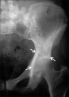
Paget's disease affecting the iliac bone demonstrated with coarsening of the bone trabeculae and cortical thickening (arrows).
Figure 14
Figure 14
Chondrosarcoma of the pelvis in (a) conventional radiography appears as an isolated osteolytic lesion (arrows), (b) Coronal T2-weighted image demonstrates a lobulated hyperintense lesion (arrowheads).

References

    1. Wolff JL, Starfield B, Anderson G. Prevalence, expenditures, and complications of multiple chronic conditions in the elderly. Arch Intern Med. 2002;162:2269–76. - PubMed
    1. Freemont AJ, Hoyland JA. Morphology, mechanisms and pathology of musculoskeletal ageing. J Pathol. 2007;211:252–9. - PubMed
    1. Cheong HW, Peh WC, Guglielmi G. Imaging of diseases of the axial and peripheral skeleton. Radiol Clin North Am. 2008;46:703–33. vi. - PubMed
    1. Kanis JA. Diagnosis of osteoporosis and assessment of fracture risk. Lancet. 2002;359:1929–36. - PubMed
    1. Gotis-Graham I, McGuigan L, Diamond T, Portek I, Quinn R, Sturgess A, et al. Sacral insufficiency fractures in the elderly. J Bone Joint Surg Br. 1994;76:882–6. - PubMed