

Prenatal Diagnosis of EEC Syndrome with "Lobster Claw" Anomaly by 3D Ultrasound
- PMID: 22919554
- PMCID: PMC3424774
- DOI: 10.4103/2156-7514.99153
Prenatal Diagnosis of EEC Syndrome with "Lobster Claw" Anomaly by 3D Ultrasound
Abstract
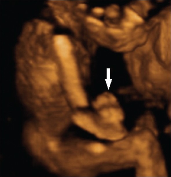
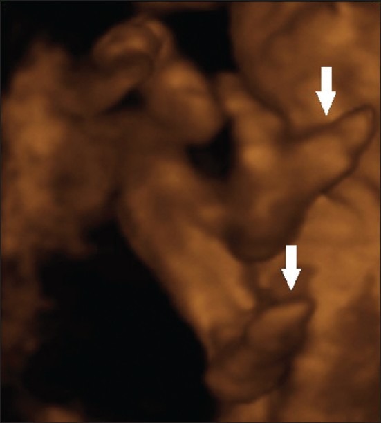
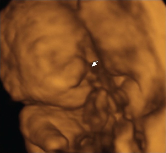
THE EEC SYNDROME IS A GENETIC ANOMALY CHARACTERIZED BY THE TRIAD: ectodermal dysplasia (development of anomalies of the structures derived from the embryonic ectodermal layer), ectrodactyly (extremities, hands and feet malformations) and cleft lip and/or palate; these malformations can be seen together or in isolation. The prenatal diagnosis can be made by two-dimensional ultrasonography (2DUS) that identifies the facial and/or limb anomalies, most characteristic being the "lobster-claw" hands. The three-dimensional ultrasonography (3DUS) provides a better analysis of the malformations than the 2DUS. A 25-year-old primigravida, had her first transvaginal ultrasonography that showed an unique fetus with crow-rump length of 47 mm with poorly defined hands and feet,. She was suspected of having sporadic form of EEC syndrome. The 2DUS performed at 19 weeks confirmed the EEC syndrome, showing a fetus with lobster-claw hands (absence of the 2(nd) and 3(rd) fingers), left foot with the absence of the 3rd toe and the right foot with syndactyly, and presence of cleft lip/palate. The 3DUS defined the anomalies much better than 2DUS including the lobster-claw hands.
Keywords: Ectodermal dysplasia; ectrodactyly; lobster claw hand; prenatal diagnosis; three-dimensional ultrasonography; two-dimensional ultrasonography.
Conflict of interest statement
Figures
References
-
- Rüdiger RA, Haase W, Passarge E. Association of ectrodactyly, ectodermal dysplasia, and cleft lip-palate. Am J Dis Child. 1970;120:160–3. - PubMed
-
- Rodini ES, Richieri-Costa A. EEC syndrome: Report on 20 new patients, clinical and genetic considerations. Am J Med Genet. 1990;37:42–53. - PubMed
-
- Janssens S, Defoort P, Vandenbroecke C, Scheffer H, Mortier G. Prune belly anomaly on prenatal ultrasound as a presenting feature of ectrodactyly-ectodermal dysplasia-clefting syndrome (EEC) Genet Couns. 2008;19:433–7. - PubMed
-
- Leung KY, MacLachlan NA, Sepulveda W. Prenatal diagnosis of ectrodactyly: The ‘lobster claw’ anomaly. Ultrasound Obstet Gynecol. 1995;6:443–6. - PubMed
Publication types
LinkOut - more resources
Full Text Sources
Miscellaneous
