. 2012 Mar 20:2:31.
doi: 10.3389/fcimb.2012.00031. eCollection 2012.

The Campylobacter jejuni CiaC virulence protein is secreted from the flagellum and delivered to the cytosol of host cells

Affiliations

The Campylobacter jejuni CiaC virulence protein is secreted from the flagellum and delivered to the cytosol of host cells

Jason M Neal-McKinney et al. Front Cell Infect Microbiol. .

Abstract

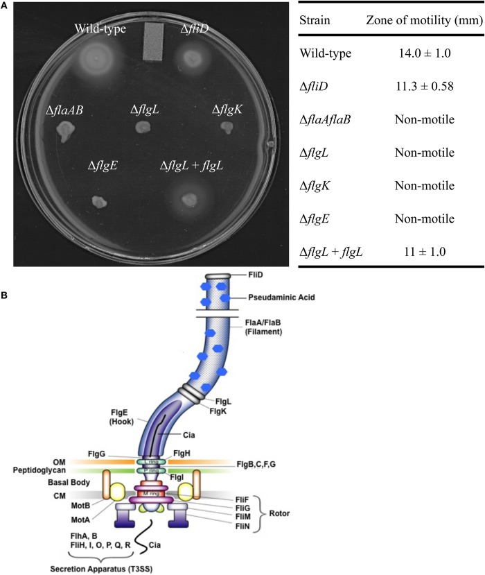
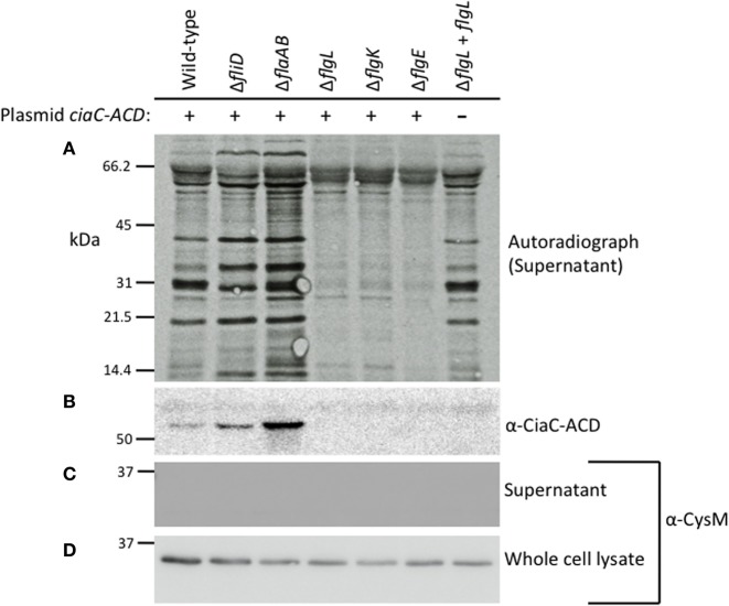
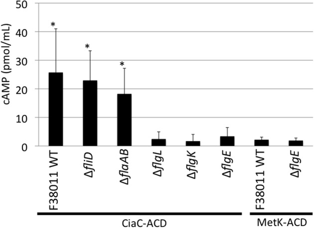
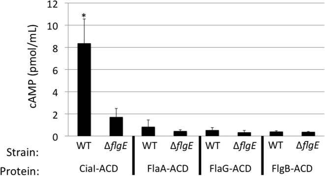
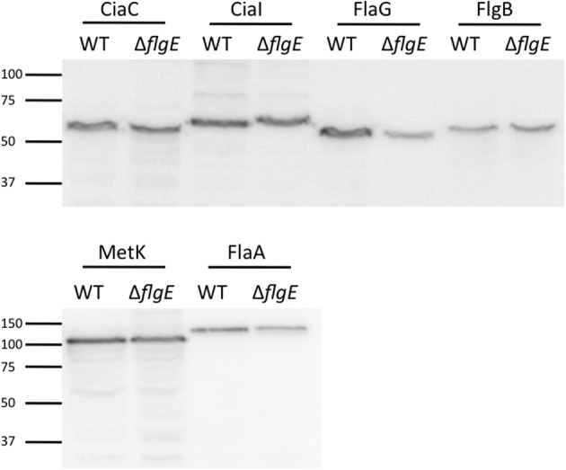
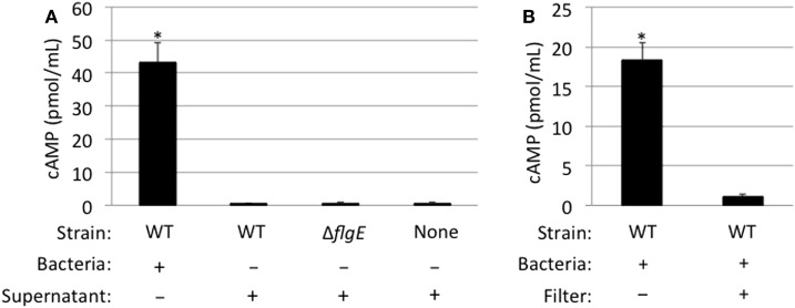
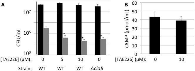
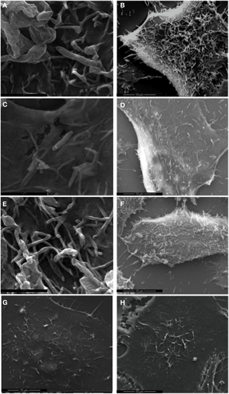
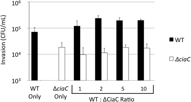
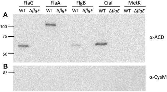
Campylobacter jejuni is a leading cause of bacterial gastroenteritis worldwide. Acute C. jejuni-mediated disease (campylobacteriosis) involves C. jejuni invasion of host epithelial cells using adhesins (e.g., CadF and FlpA) and secreted proteins [e.g., the Campylobacter invasion antigens (Cia)]. The genes encoding the Cia proteins are up-regulated upon co-culture of C. jejuni with epithelial cells. One of the Cia proteins, CiaC, is required for maximal invasion of host cells by C. jejuni. Previous work has also revealed that CiaC is, in part, responsible for host cell cytoskeletal rearrangements that result in membrane ruffling. This study was performed to test the hypothesis that CiaC is delivered to the cytosol of host cells. To detect the delivery of CiaC into cultured epithelial cells, we used the adenylate cyclase domain (ACD) of Bordetella pertussis CyaA as a reporter. In this study, we found that export and delivery of the C. jejuni Cia proteins into human INT 407 epithelial cells required a functional flagellar hook complex composed of FlgE, FlgK, and FlgL. Assays performed with bacterial culture supernatants supported the hypothesis that CiaC delivery requires bacteria-host cell contact. We also found that CiaC was delivered to host cells by cell-associated (bound) bacteria, as judged by experiments performed with inhibitors that specifically target the cell signaling pathways utilized by C. jejuni for cell invasion. Interestingly, the C. jejuni flgL mutant, which is incapable of exporting and delivering the Cia proteins, did not induce INT 407 cell membrane ruffles. Complementation of the flgL mutant with plasmid-encoded flgL restored the motility and membrane ruffling. These data support the hypothesis that the C. jejuni Cia proteins, which are exported from the flagellum, are delivered to the cytosol of host cells.

Keywords: T3SS; adenylate cyclase; effector proteins; flagellum; membrane ruffling.


Figures

Figure 1
Figure 1
Motility of the C. jejuni flagellar mutants. Motility assays were performed using C. jejuni F38011 wild-type strain, the isogenic fliD, flaAflaB, flgL, flgK, and flgE mutants, and the flgL mutant complemented with flgL (A). The table contains zones of motility ± one standard deviation obtained from a quantitative motility assay performed in triplicate. The zone of motility was determined by measuring from the edge of the inoculum spot to the edge of growth. A diagram of the C. jejuni flagellum is also included, and was adopted from [Larson et al. (2008)] (B). OM, outermembrane; CM, cytoplasmic membrane.
Figure 2
Figure 2
The Cia proteins are secreted from the C. jejuni F38011 wild-type strain and fliD and flaAflaB mutants, but not flgL, flgK, and flgE mutants. The C. jejuni F38011 wild-type strain and isogenic fliD, flaAB, flgL, flgK, and flgE mutants were assayed for secretion of the Cia proteins as well as for secretion of plasmid-encoded CiaC-ACD. Newly synthesized proteins were labeled with [35S]-methionine and Cia secretion was stimulated by the addition of 1% FBS. Proteins in the supernatants were concentrated 10-fold, separated by SDS-PAGE, transferred to PVDF membranes, and exposed to X-ray film (A) or probed with an anti-ACD antibody (B). Faint bands present in all lanes represent bovine serum albumin (66.5 kDa) and degradation products. The molecular mass of the pre-stained protein standards are listed in kDa. Supernatants (C) and whole-cell lysates (D) were probed with anti-CysM antibody to confirm that cell lysis did not occur.
Figure 3
Figure 3
The CiaC virulence proteins is delivered to the cytosol of INT 407 cells from the C. jejuni F38011 wild-type strain, fliD mutant, and flaAflaB mutant, but not flgL, flgK, and flgE mutants. Delivery of the CiaC-ACD fusion protein from the cytosol of C. jejuni to the cytosol of INT 407 cells was assayed in the C. jejuni F38011 wild-type strain and isogenic fliD, flaAflaB, flgL, flgK, and flgE mutants, and delivery of MetK-ACD was assayed in the C. jejuni F38011 wild-type strain and flgE mutant. The concentration of cAMP within the INT 407 cells was assayed 30 min post-infection. The error bars represent one standard deviation from mean. The asterisk indicates values that are significantly different from the flgE mutant harboring ciaC-ACD as judged by the Student's t-test (P < 0.05). Data from two biological replicates were combined.
Figure 4
Figure 4
CiaI is delivered from the C. jejuni F38011 wild-type strain to INT 407 cells. Delivery of the CiaI-ACD, FlaA-ACD, FlaG-ACD, and FlgB-ACD fusion proteins was assayed in the C. jejuni F38011 wild-type strain and isogenic flgE mutant. INT 407 cells were inoculated with the C. jejuni F38011 wild-type strain and flgE mutant harboring the various ACD plasmids. The concentration of cAMP within the INT 407 cells was assayed 30 min post-infection. The error bars represent one standard deviation from mean. The asterisk indicates that the amount of cAMP produced in the wild-type strain was significantly greater than the value obtained from the flgE mutant, as judged by the Student's t-test (P < 0.05).
Figure 5
Figure 5
Immunoblot using anti-ACD antibody reveals equivalent protein synthesis of ACD fusion proteins in C. jejuni F38011 wild-type strain and flgE mutant. The amount of ACD fusion proteins produced in the C. jejuni F38011 wild-type strain and isogenic flgE mutant was examined by immunoblot. Whole cell lysates were prepared from bacteria grown overnight in MH broth supplemented with 0.01% deoxycholate, transferred to PVDF membranes, and probed with an anti-ACD antibody. The molecular mass of the pre-stained protein standards are listed in kDa.
Figure 6
Figure 6
CiaC-ACD delivery into INT 407 cells requires bacteria-host cell contact. The delivery of CiaC-ACD into INT 407 cells assayed using whole bacteria and CiaC-ACD containing supernatants (A), as well as a filter to block physical contact of bacteria with INT 407 cells (B). (A) Cell-free supernatants of C. jejuni F38011 wild-type strain and isogenic flgE mutant were harvested from bacteria incubated in MEM supplemented with 1% FBS for 1 h. The C. jejuni F38011 strain harboring the ciaC-ACD plasmid or cell-free supernatants collected from this strain were added to INT 407 cells and cAMP was measured 30 min post-inoculation. (B) A delivery assay was performed in the presence and absence of a 0.2 μm pore filter. To accommodate the 0.2 μm pore filter, six-well tissue culture plates (rather than 24-well) were used. The value shown for C. jejuni F38011 wild-type strain was normalized by subtracting the background cAMP value obtained for the flgE mutant, with or without a 0.2 μm pore filter. The error bars represent one standard deviation from mean. The asterisk indicates the amount of cAMP produced in the wild-type strain was significantly greater than the value obtained from the flgE mutant as judged by the Student's t-test (P < 0.05)
Figure 7
Figure 7
CiaC-ACD is delivered into INT 407 cells by extracellular bacteria. A bacterial invasion assay (A) and delivery assay (B) were performed in the presence of TAE226, an inhibitor of Focal Adhesion Kinase. Focal adhesion kinase is required for maximal invasion of host cells by C. jejuni. (A) The gentamicin protection assay was used to determine the number of cells of C. jejuni F38011 wild-type strain and its isogenic ciaB mutant internalized in the presence of 0, 5, or 10 μM TAE226. Gentamicin was added 3 h post-infection to kill extracellular bacteria. The INT 407 cells were then lysed and the bacteria were enumerated by serial dilution and plating. Shown are the number of cell-associated bacteria (black bars) and internalized bacteria (gray bars). The number of bacteria internalized in the presence of TAE226 was significantly reduced, when compared to C. jejuni infected cells in the absence of inhibitor, as judged by the Student's t-test (P < 0.05). (B) Delivery of the CiaC-ACD fusion protein was assayed at 30 min post-infection in the presence of 0 or 10 μM TAE226. The values shown were normalized by subtracting the background cAMP value obtained for the flgE mutant (i.e., Cia secretion negative) containing each ACD fusion construct from the values obtained for the wild-type strain. The error bars represent one standard deviation from the mean. The amount of cAMP produced in the presence of TAE226 was not significantly different from the value obtained in the absence of TAE226, as judged by the Student's t-test (P < 0.05). Cells were treated with methanol (vehicle control), and trypan blue staining was performed to confirm that the solvent used for TAE226 did not affect cell viability (not shown).
Figure 8
Figure 8
C. jejuni F38011 wild-type induces membrane ruffling. INT 407 cells were infected with the C. jejuni F38011 wild-type strain (A and B), flgL mutant (C and D), flgL mutant harboring a plasmid containing native flgL (E and F), cell-free supernatants containing the Cia proteins (G), or were uninfected (H). The cells were fixed and removed at 15 min post-infection, and visualized by SEM at 50,000× (A, C, and E) and 7000× (B, D, F, G, and H) magnification.
Figure 9
Figure 9
Cia delivery by the C. jejuni F38011 wild-type strain cannot rescue the invasion of a ciaC mutant. A bacterial invasion assay was performed using the C. jejuni F38011 wild-type strain, the isogenic ciaC mutant, or a mixture of both strains. The numbers of internalized C. jejuni F38011 wild-type (black bars) and ciaC mutant (white bars) bacteria were determined at 3 h post-infection using the gentamicin protection assay. The numbers below the graph indicate the quantity of C. jejuni F38011 wild-type and ciaC mutant bacteria in each well, in units of 0.03 OD540. The error bars represent one standard deviation. The number of C. jejuniciaC mutant bacteria internalized did not change in response to increasing amounts of wild-type bacteria, as judged by the Student's t-test (P < 0.05).
Figure A1
Figure A1
Complementation of the C. jejuni flgL mutant with a plasmid harboring flgL restores the flagellar filament. C. jejuni wild-type strain, the flgL mutant, and flgL mutant harboring plasmid-encoded flgL were examined by TEM. C. jejuni were spotted onto formvar coated copper grids, stained with 2% phosphotungstic acid, and visualized at 6500× magnification. The scale bar represents one micron.
Figure A2
Figure A2
C. jejuni 81116 and 81–176 flaAflaB mutants secrete the Cia proteins. Secretion of the Cia proteins from 81116 and 81–176 wild-type strains and isogenic flaAflaB mutants was stimulated by incubation with 1% FBS, and the secreted proteins were analyzed by autoradiograph.
Figure A3
Figure A3
FlaG, FlaA, FlgB, and CiaI are secreted through the flagellar T3SS. Secretion of the FlaG-, FlaA-, FlgB-, CiaI-, and MetK-ACD fusion proteins from the C. jejuni wild-type strain and flgE mutant was examined. The C. jejuni strains were transformed with the various ACD plasmids, and Cia secretion was stimulated by the addition of FBS. After 3 h, the proteins in the supernatant fractions were precipitated and an immunoblot was performed using an anti-ACD (A) and anti-CysM (B) antibodies. MetK-ACD was included as a negative control, as it is not secreted from C. jejuni. The molecular mass of the pre-stained protein standards are listed in kDa.

References

    1. Ashgar S. S., Oldfield N. J., Wooldridge K. G., Jones M. A., Irving G. J., Turner D. P., Ala'Aldeen D. A. (2007). CapA, an autotransporter protein of Campylobacter jejuni, mediates association with human epithelial cells and colonization of the chicken gut. J. Bacteriol. 189, 1856–1865 10.1128/JB.01427-06 - DOI - PMC - PubMed
    1. Badea L., Beatson S. A., Kaparakis M., Ferrero R. L., Hartland E. L. (2009). Secretion of flagellin by the LEE-encoded type III secretion system of enteropathogenic Escherichia coli. BMC Microbiol. 9, 30 10.1186/1471-2180-9-30 - DOI - PMC - PubMed
    1. Bardill J. P., Miller J. L., Vogel J. P. (2005). IcmS-dependent translocation of SdeA into macrophages by the Legionella pneumophila type IV secretion system. Mol. Microbiol. 56, 90–103 10.1111/j.1365-2958.2005.04539.x - DOI - PubMed
    1. Black R. E., Levine M. M., Clements M. L., Hughes T. P., Blaser M. J. (1988). Experimental Campylobacter jejuni infection in humans. J. Infect. Dis. 157, 472–479 10.1093/infdis/157.3.472 - DOI - PubMed
    1. Buelow D. R., Christensen J. E., Neal-Mckinney J. M., Konkel M. E. (2010). Campylobacter jejuni survival within human epithelial cells is enhanced by the secreted protein CiaI. Mol. Microbiol. 80, 1296–1312 10.1111/j.1365-2958.2011.07645.x - DOI - PMC - PubMed

Publication types

LinkOut - more resources