. 2012 May 11:2:61.
doi: 10.3389/fcimb.2012.00061. eCollection 2012.

O-antigen and virulence profiling of shiga toxin-producing Escherichia coli by a rapid and cost-effective DNA microarray colorimetric method

Affiliations

O-antigen and virulence profiling of shiga toxin-producing Escherichia coli by a rapid and cost-effective DNA microarray colorimetric method

Beatriz Quiñones et al. Front Cell Infect Microbiol. .

Abstract

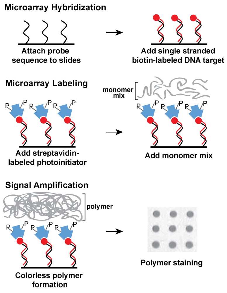
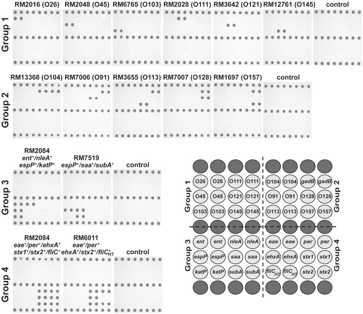
Shiga toxin-producing Escherichia coli (STEC) is a leading cause of foodborne illness worldwide. The present study developed the use of DNA microarrays with the ampliPHOX colorimetric method to rapidly detect and genotype STEC strains. A low-density 30-mer oligonucleotide DNA microarray was designed to target O-antigen gene clusters of 11 E. coli serogroups (O26, O45, O91, O103, O104, O111, O113, O121, O128, O145, and O157) that have been associated with the majority of STEC infections. In addition, the DNA microarray targeted 11 virulence genes, encoding adhesins, cytotoxins, proteases, and receptor proteins, which have been implicated in conferring increased ability to cause disease for STEC. Results from the validation experiments demonstrated that this microarray-based colorimetric method allowed for a rapid and accurate genotyping of STEC reference strains from environmental and clinical sources and from distinct geographical locations. Positive hybridization signals were detected only for probes targeting serotype and virulence genes known to be present in the STEC reference strains. Quantification analysis indicated that the mean pixel intensities of the signal for probes targeting O-antigen or virulence genes were at least three times higher when compared to the background. Furthermore, this microarray-based colorimetric method was then employed to genotype a group of E. coli isolates from watershed sediment and animal fecal samples that were collected from an important region for leafy-vegetable production in the central coast of California. The results indicated an accurate identification of O-type and virulence genes in the tested isolates and confirmed that the ampliPHOX colorimetric method with low-density DNA microarrays enabled a fast assessment of the virulence potential of STEC using low-cost reagents and instrumentation.

Keywords: DNA microarrays; Escherichia coli; STEC; Shiga toxin; ampliPHOX; foodborne pathogen; genotyping; photopolymerization.


Figures

Figure 1
Figure 1
Schematic diagram of ampliPHOX colorimetric method with DNA microarrays. First, DNA microarrays are hybridized with DNA target labeled with biotin (red circles). Second, the microarray is labeled with a photoinitiator (letter P) that is conjugated to streptavidin (blue polygons). Third, a short light-initiated polymerization reaction results in a colorless polymer localized exclusively where the probe and target sequences hybridized on the microarray. Polymer formation is visualized after a quick staining step.
Figure 2
Figure 2
Genotyping Shiga toxin-producing Escherichia coli (STEC) with low-density DNA microarrays and the ampliPHOX colorimetric method. STEC reference strains from clinical or environmental sources with different genotypes (Table 1) were analyzed by 30-mer oligonucleotide DNA microarrays and the ampliPHOX colorimetric method. The panels, shown under strain designations, illustrate polymer formation on spotted probes for microarrays hybridized with amplification reactions testing a particular STEC reference strain or controls lacking a DNA template. The array layout shows the duplicate spots with probes targeting the O-antigen and virulence genes that are represented in groups 1–4. Biotinylated probes spotted along the top, middle and bottom rows (dark gray) of the array layout correspond to positive controls for polymer formation.

References

    1. Anjum M. F., Mafura M., Slickers P., Ballmer K., Kuhnert P., Woodward M. J., Ehricht R. (2007). Pathotyping Escherichia coli by using miniaturized DNA microarrays. Appl. Environ. Microbiol. 73, 5692–569710.1128/AEM.00419-07 - DOI - PMC - PubMed
    1. Ballmer K., Korczak B. M., Kuhnert P., Slickers P., Ehricht R., Hachler H. (2007). Fast DNA serotyping of Escherichia coli by use of an oligonucleotide microarray. J. Clin. Microbiol. 45, 370–37910.1128/JCM.01361-06 - DOI - PMC - PubMed
    1. Bettelheim K. A. (2007). The non-O157 Shiga-toxigenic (verocytotoxigenic) Escherichia coli; under-rated pathogens. Crit. Rev. Microbiol. 33, 67–8710.1080/10408410601172172 - DOI - PubMed
    1. Boissinot K., Huletsky A., Peytavi R., Turcotte S., Veillette V., Boissinot M., Picard F. J., Martel E. A., Bergeron M. G. (2007). Rapid exonuclease digestion of PCR-amplified targets for improved microarray hybridization. Clin. Chem. 53, 2020–202310.1373/clinchem.2007.091157 - DOI - PubMed
    1. Bolton D. J. (2011). Verocytotoxigenic (Shiga toxin-producing) Escherichia coli: virulence factors and pathogenicity in the farm to fork paradigm. Foodborne Pathog. Dis. 8, 357–36510.1089/fpd.2010.0699 - DOI - PubMed

Publication types