

The early interaction of Leishmania with macrophages and dendritic cells and its influence on the host immune response
- PMID: 22919674
- PMCID: PMC3417671
- DOI: 10.3389/fcimb.2012.00083
The early interaction of Leishmania with macrophages and dendritic cells and its influence on the host immune response
Abstract
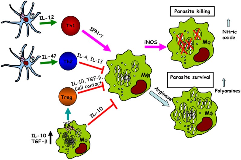
The complicated interactions between Leishmania and the host antigen-presenting cells (APCs) have fundamental effects on the final outcome of the disease. Two major APCs, macrophages and dendritic cells (DCs), play critical roles in mediating resistance and susceptibility during Leishmania infection. Macrophages are the primary resident cell for Leishmania: they phagocytose and permit parasite proliferation. However, these cells are also the major effector cells to eliminate infection. The effective clearance of parasites by macrophages depends on activation of appropriate immune response, which is usually initiated by DCs. Here, we review the early interaction of APCs with Leishmania parasites and how these interactions profoundly impact on the ensuing adaptive immune response. We also discuss how the current knowledge will allow further refinement of our understanding of the interplay between Leishmania and its hosts that leads to resistance or susceptibility.
Keywords: cytokines; dendritic cells; innate immunity; macrophages; parasitic-protozoan; rodents.
Figures
References
-
- Abou Fakher F. H., Rachinel N., Klimczak M., Louis J., Doyen N. (2009). TLR9-dependent activation of dendritic cells by DNA from Leishmania major favors Th1 cell development and the resolution of lesions. J. Immunol. 182, 1386–1396 - PubMed
-
- Balaraman S., Singh V. K., Tewary P., Madhubala R. (2005). Leishmania lipophosphoglycan activates the transcription factor activating protein 1 in J774A.1 macrophages through the extracellular signal-related kinase (ERK) and p38 mitogen-activated protein kinase. Mol. Biochem. Parasitol. 139, 117–127 10.1016/j.molbiopara.2004.10.006 - DOI - PubMed
Publication types
MeSH terms
LinkOut - more resources
Full Text Sources
Medical
Miscellaneous