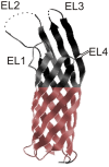
Yersinia pestis Ail: multiple roles of a single protein
- PMID: 22919692
- PMCID: PMC3417512
- DOI: 10.3389/fcimb.2012.00103
Yersinia pestis Ail: multiple roles of a single protein
Abstract
Yersinia pestis is one of the most virulent bacteria identified. It is the causative agent of plague-a systemic disease that has claimed millions of human lives throughout history. Y. pestis survival in insect and mammalian host species requires fine-tuning to sense and respond to varying environmental cues. Multiple Y. pestis attributes participate in this process and contribute to its pathogenicity and highly efficient transmission between hosts. These include factors inherited from its enteric predecessors; Y. enterocolitica and Y. pseudotuberculosis, as well as phenotypes acquired or lost during Y. pestis speciation. Representatives of a large Enterobacteriaceae Ail/OmpX/PagC/Lom family of outer membrane proteins (OMPs) are found in the genomes of all pathogenic Yersiniae. This review describes the current knowledge regarding the role of Ail in Y. pestis pathogenesis and virulence. The pronounced role of Ail in the following areas are discussed (1) inhibition of the bactericidal properties of complement, (2) attachment and Yersinia outer proteins (Yop) delivery to host tissue, (3) prevention of PMNL recruitment to the lymph nodes, and (4) inhibition of the inflammatory response. Finally, Ail homologs in Y. enterocolitica and Y. pseudotuberculosis are compared to illustrate differences that may have contributed to the drastic bacterial lifestyle change that shifted Y. pestis from an enteric to a vector-born systemic pathogen.
Keywords: Ail; OmpX; T3SS; Yersinia pestis; adhesion; invasion; serum resistance; virulence.
Figures


Similar articles
-
Ail provides multiple mechanisms of serum resistance to Yersinia pestis.Mol Microbiol. 2019 Jan;111(1):82-95. doi: 10.1111/mmi.14140. Epub 2018 Oct 26. Mol Microbiol. 2019. PMID: 30260060 Free PMC article.
-
Ail proteins of Yersinia pestis and Y. pseudotuberculosis have different cell binding and invasion activities.PLoS One. 2013 Dec 27;8(12):e83621. doi: 10.1371/journal.pone.0083621. eCollection 2013. PLoS One. 2013. PMID: 24386237 Free PMC article.
-
Phenotypic characterization of OmpX, an Ail homologue of Yersinia pestis KIM.Microbiology (Reading). 2007 Sep;153(Pt 9):2941-2951. doi: 10.1099/mic.0.2006/005694-0. Microbiology (Reading). 2007. PMID: 17768237
-
Contributions of Yersinia pestis outer membrane protein Ail to plague pathogenesis.Curr Opin Infect Dis. 2022 Jun 1;35(3):188-195. doi: 10.1097/QCO.0000000000000830. Curr Opin Infect Dis. 2022. PMID: 35665712 Free PMC article. Review.
-
Adhesins of human pathogens from the genus Yersinia.Adv Exp Med Biol. 2011;715:1-15. doi: 10.1007/978-94-007-0940-9_1. Adv Exp Med Biol. 2011. PMID: 21557054 Review.
Cited by
-
Unraveling neutrophil- Yersinia interactions during tissue infection.F1000Res. 2019 Jul 11;8:F1000 Faculty Rev-1046. doi: 10.12688/f1000research.18940.1. eCollection 2019. F1000Res. 2019. PMID: 31327994 Free PMC article. Review.
-
Mutually constructive roles of Ail and LPS in Yersinia pestis serum survival.Mol Microbiol. 2020 Sep;114(3):510-520. doi: 10.1111/mmi.14530. Epub 2020 Jun 25. Mol Microbiol. 2020. PMID: 32462782 Free PMC article.
-
Environmental Regulation of Yersinia Pathophysiology.Front Cell Infect Microbiol. 2016 Mar 2;6:25. doi: 10.3389/fcimb.2016.00025. eCollection 2016. Front Cell Infect Microbiol. 2016. PMID: 26973818 Free PMC article. Review.
-
Yersinia pestis Δail Mutants Are Not Susceptible to Human Complement Bactericidal Activity in the Flea.Appl Environ Microbiol. 2023 Feb 28;89(2):e0124422. doi: 10.1128/aem.01244-22. Epub 2023 Feb 6. Appl Environ Microbiol. 2023. PMID: 36744930 Free PMC article.
-
Transport proteins promoting Escherichia coli pathogenesis.Microb Pathog. 2014 Jun-Jul;71-72:41-55. doi: 10.1016/j.micpath.2014.03.008. Epub 2014 Apr 18. Microb Pathog. 2014. PMID: 24747185 Free PMC article.
References
-
- Achtman M., Morelli G., Zhu P., Wirth T., Diehl I., Kusecek B., Vogler A. J., Wagner D. M., Allender C. J., Easterday W. R., Chenal-Francisque V., Worsham P., Thomson N. R., Parkhill J., Lindler L. E., Carniel E., Keim P. (2004). Microevolution and history of the plague bacillus, Yersinia pestis. Proc. Natl. Acad. Sci. U.S.A. 101, 17837–17842 10.1073/pnas.0408026101 - DOI - PMC - PubMed
-
- Anisimov A. P., Dentovskaya S. V., Titareva G. M., Bakhteeva I. V., Shaikhutdinova R. Z., Balakhonov S. V., Lindner B., Kocharova N. A., Senchenkova S. N., Holst O., Pier G. B., Knirel Y. A. (2005). Intraspecies and temperature-dependent variations in susceptibility of Yersinia pestis to the bactericidal action of serum and to polymyxin B. Infect. Immun. 73, 7324–7331 10.1128/IAI.73.11.7324-7331.2005 - DOI - PMC - PubMed
Publication types
MeSH terms
Substances
Grants and funding
LinkOut - more resources
Full Text Sources
Molecular Biology Databases

