. 2012 Nov 15;448(1):73-82.
doi: 10.1042/BJ20120537.

Generation and characterization of non-competitive furin-inhibiting nanobodies

Affiliations

Generation and characterization of non-competitive furin-inhibiting nanobodies

Jingjing Zhu et al. Biochem J. .

Abstract

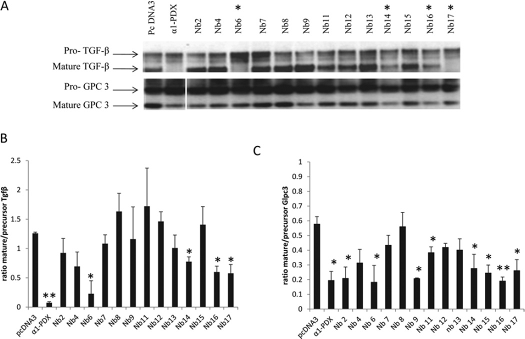
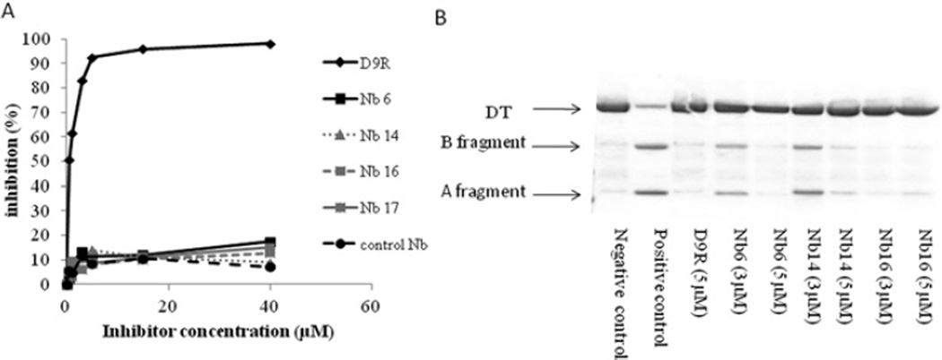
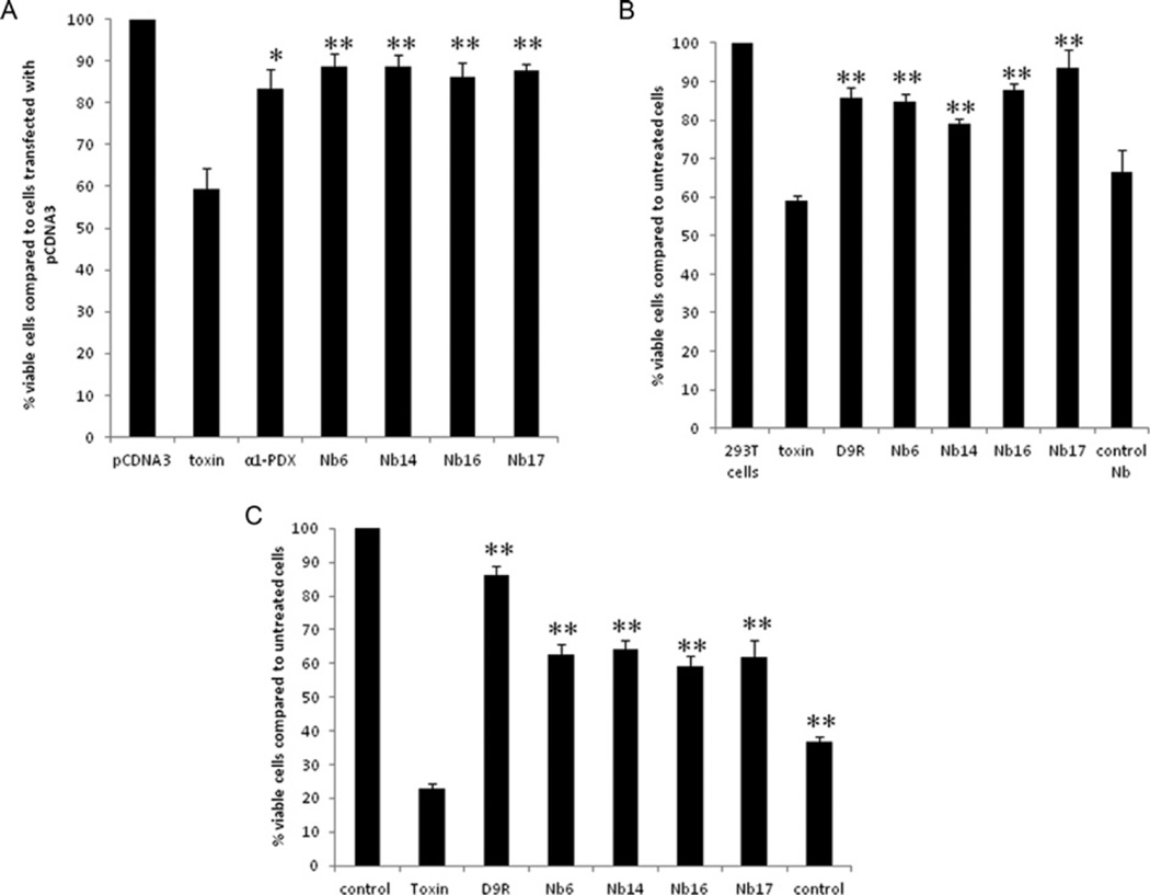
The PC (proprotein convertase) furin cleaves a large variety of proproteins and hence plays a major role in many pathologies. Therefore furin inhibition might be a good strategy for therapeutic intervention, and several furin inhibitors have been generated, although none are entirely furin-specific. To reduce potential side effects caused by cross-reactivity with other proteases, dromedary heavy-chain-derived nanobodies against catalytically active furin were developed as specific furin inhibitors. The nanobodies bound only to furin but not to other PCs. Upon overexpression in cell lines, they inhibited the cleavage of two different furin substrates, TGFβ (transforming growth factor β) and GPC3 (glypican 3). Purified nanobodies could inhibit the cleavage of diphtheria toxin into its enzymatically active A fragment, but did not inhibit cleavage of a small synthetic peptide-based substrate, suggesting a mode-of-action based on steric hindrance. The dissociation constant of purified nanobody 14 is in the nanomolar range. The nanobodies were non-competitive inhibitors with an inhibitory constant in the micromolar range as demonstrated by Dixon plot. Furthermore, anti-furin nanobodies could protect HEK (human embryonic kidney)-293T cells from diphtheria-toxin-induced cytotoxicity as efficiently as the PC inhibitor nona-D-arginine. In conclusion, these antibody-based single-domain nanobodies represent the first generation of highly specific non-competitive furin inhibitors.


Figures

Figure 1
Figure 1. Co-immunoprecipitation of human furin with the different nanobodies
RPE.40 cells were transfected with human furin with or without expression vectors encoding the different nanobodies tagged with HA and immunoprecipitation was performed using an anti-HA antibody. Subsequently, Western blot analysis was performed using the anti-furin antibody MON152. Lanes 1, beads without anti-HA antibody; lanes 2, beads with anti-HA antibody; lanes 3, total cell lysate. Untransfected cells were included as a negative control in the last lane (−)
Figure 2
Figure 2. Four different nanobodies inhibit the cleavage of the furin substrates TGFβ and GPC3
HEK-293T cells were transfected with an expression vector encoding TGFβ or GPC3 together with empty vector, α1-PDX or an expression vector encoding the different nanobodies. (A) The inhibition of the furin-mediated cleavage of those substrates was analysed by Western blotting. Nb6, Nb14, Nb16 and Nb17 inhibited the cleavage of the two furin substrates (indicated with *). (B) The ratio of mature/precursor TGFβ was calculated from three different experiments using ImageJ software. Results are represented as means ± S.E.M. (n =3) *P ≤0.05, **P ≤0.005. (C) The ratio of mature/precursor GPC3 was calculated from three different experiments using ImageJ software. Results are represented as means ± S.E.M. (n =3) *P ≤0.05, **P ≤0.005.
Figure 3
Figure 3. Nanobodies bind to mouse and human furin, but not to the other PC family members
(A) RPE.40 cells were transfected with furin cDNA or cDNAs encoding closely related PCs with or without cDNAs encoding the different nanobodies and immunoprecipitation was performed using an anti-HA antibody. Only mouse and human furin, but not family members, co-immunoprecipitated with Nb6, Nb14, Nb16 and Nb17, indicating their specificity for furin. A representative image for Nb14 is shown. Lane 1, beads without anti-HA antibody; lane 2, beads with anti-HA antibody; lane 3, total cell lysate. (B) RPE.40 cells were transfected with a construct encoding the mutant RVRTKR of renin-2 and furin cDNA or cDNAs encoding closely related PCs together with the empty pcDNA3 vector (lane 1), α1-PDX (lane 2) or cDNA encoding Nb6 (lane 3). Subsequently, Western blot analysis was performed using the anti-renin antibody. h, human; m, mouse.
Figure 4
Figure 4. Purified nanobodies cannot inhibit the cleavage of the small pyr-Arg-Thr-Lys-Arg-AMC substrate, but do inhibit the cleavage of diphtheria toxin
(A) Purified Nb6, Nb14, Nb16 and Nb17 and a control nanobody cannot inhibit furin-mediated cleavage of pyr-Arg-Thr-Lys-Arg-AMC, as shown by a fluorimetric assay. In contrast, the well-characterized furin inhibitor D9R effectively inhibits the cleavage of this substrate. (B) D9R and purified Nb6, Nb14 and Nb16 inhibit the furin-mediated cleavage of diphtheria toxin (DT), as shown by Coomassie Brilliant Blue staining.
Figure 5
Figure 5. The nanobodies inhibit diphtheria-toxin-mediated cytotoxicity as efficiently as α1-PDX and D9R
(A) HEK-293T cells were transfected with empty vector, an expression vector encoding α1-PDX or the different nanobodies 24 h before exposure to diphtheria toxin. Then, 3 h after adding diphtheria toxin, cell viability was assessed by the MTT assay. The nanobodies protected the cells from cytotoxicity as efficiently as the well-characterized furin inhibitor α1-PDX. The viability of cells treated with the diphtheria toxin together with α1-PDX or the different nanobodies is significantly higher when compared with cells only treated with the diphtheria toxin. (B) At 2 h prior to the exposure to diphtheria toxin, HEK-293T cells were incubated with 10 µM of purified nanobodies or with D9R. Then 1.5 h after adding the diphtheria toxin, cell viability was assessed by the MTT assay. The nanobodies targeting furin significantly protected the cells from cytotoxicity; protection was as efficient as the well-characterized furin inhibitor D9R, whereas control nanobodies which do not target furin did not inhibit cell toxicity. (C) At 30 min prior to the exposure to anthrax toxin (200 ng/ml PA and 400 ng/ml LF), RAW cells were incubated with 20 µM purified nanobodies or with 10 µM D9R. Then, 1.5 h after adding the toxins cell viablility was assayed using the MTT assay. Nanobodies targeting furin significantly protected the cells from cytotoxicity although less efficient than D9R, whereas control nanobodies which do not target furin only showed a mild effect on the cell toxicity. Results are presented as means ± S.E.M. (n =3) *P ≤0.05, **P ≤0.005.
Figure 6
Figure 6. Antibody uptake experiments
Antibody uptake experiments (30 min at 4°C, 15 min at 37°C) using the anti-HA antibody to detect the HA-tagged nanobodies (A) and anti-FLAG antibody M2 (B) were performed on HEK-293T cells transfected with FLAG-tagged furin.
Figure 7
Figure 7. The nanobodies are non-competitive inhibitors with Ki values in the micromolar range
(AD) Dixon plot for inhibition of furin-mediated cleavage of diphtheria toxin by Nb6 (A), Nb14 (B), Nb16 (C) and Nb17 (D). The reciprocal velocity is plotted against the inhibitor concentration. The trendlines drawn for each substrate concentration intersect in a single point on the x-axis, indicating non-competitive inhibition. (E) Dixon plot for inhibition of furin-mediated cleavage of diphtheria toxin by D9R. The reciprocal velocity is plotted against the inhibitor concentration. The trendlines drawn for each concentration of substrate intersect in a single point above the x-axis, indicating competitive inhibition. (F) Single cycle kinetic analysis for Nb14-furin interaction. Nb14 (50, 150, 300, 450 and 600 nM) was injected consecutively over the immobilized furin. Binding of Nb14 (in RU) is shown in grey as a function of time. The start and end of the injections are indicated with closed and open arrows respectively. The fitted model is indicated with a broken black line. One out of three experiments is shown as a representative Figure.

References

    1. Taylor NA, Van De Ven WJ, Creemers JW. Curbing activation: proprotein convertases in homeostasis and pathology. FASEB J. 2003;17:1215–1227. - PubMed
    1. Creemers JW, Khatib AM. Knock-out mouse models of proprotein convertases: unique functions or redundancy? Front. Biosci. 2008;13:4960–4971. - PubMed
    1. Roebroek AJ, Umans L, Pauli IG, Robertson EJ, van Leuven F, Van de Ven WJ, Constam DB. Failure of ventral closure and axial rotation in embryos lacking the proprotein convertase Furin. Development. 1998;125:4863–4876. - PubMed
    1. Roebroek AJ, Taylor NA, Louagie E, Pauli I, Smeijers L, Snellinx A, Lauwers A, Van de Ven WJ, Hartmann D, Creemers JW. Limited redundancy of the proprotein convertase furin in mouse liver. J. Biol. Chem. 2004;279:53442–53450. - PubMed
    1. De Vos L, Declercq J, Rosas GG, Van Damme B, Roebroek A, Vermorken F, Ceuppens J, van de Ven W, Creemers J. MMTV-cre-mediated fur inactivation concomitant with PLAG1 proto-oncogene activation delays salivary gland tumorigenesis in mice. Int. J. Oncol. 2008;32:1073–1083. - PubMed

Publication types

MeSH terms