. 2012 Aug 23;75(4):633-47.
doi: 10.1016/j.neuron.2012.06.021.

c-Jun reprograms Schwann cells of injured nerves to generate a repair cell essential for regeneration

Affiliations

c-Jun reprograms Schwann cells of injured nerves to generate a repair cell essential for regeneration

Peter J Arthur-Farraj et al. Neuron. .

Abstract

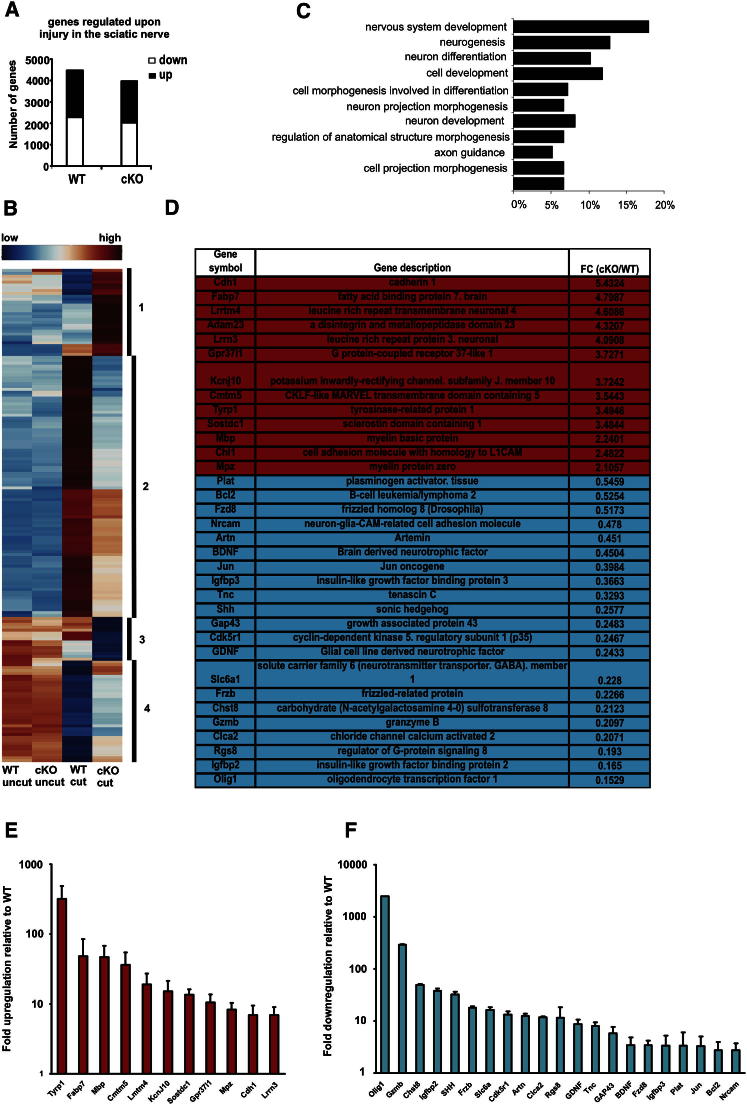
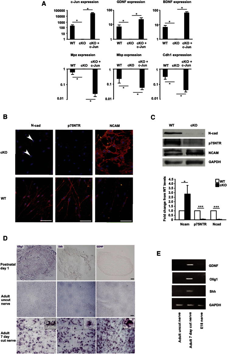
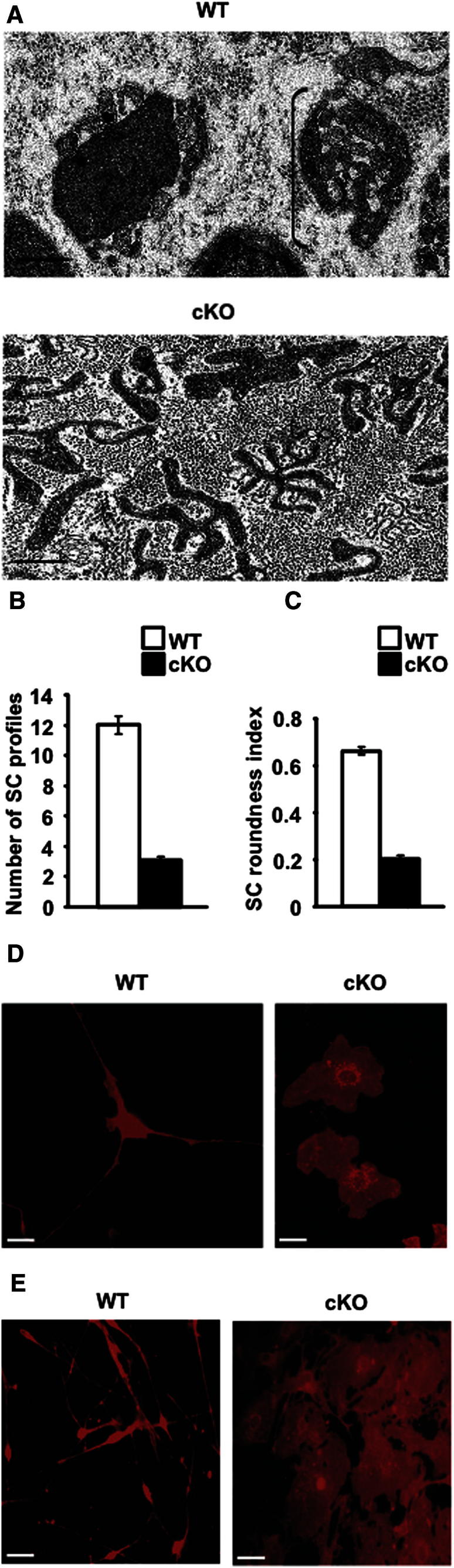
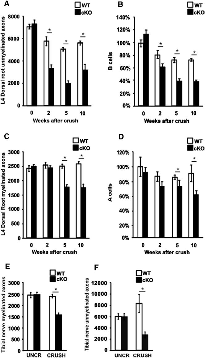
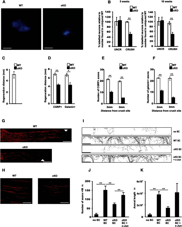
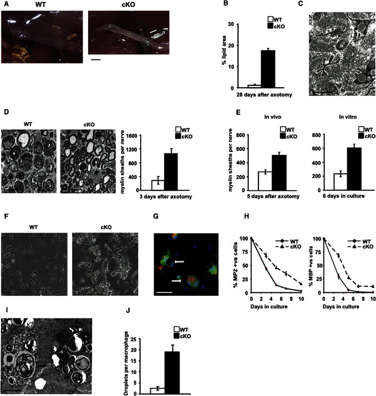
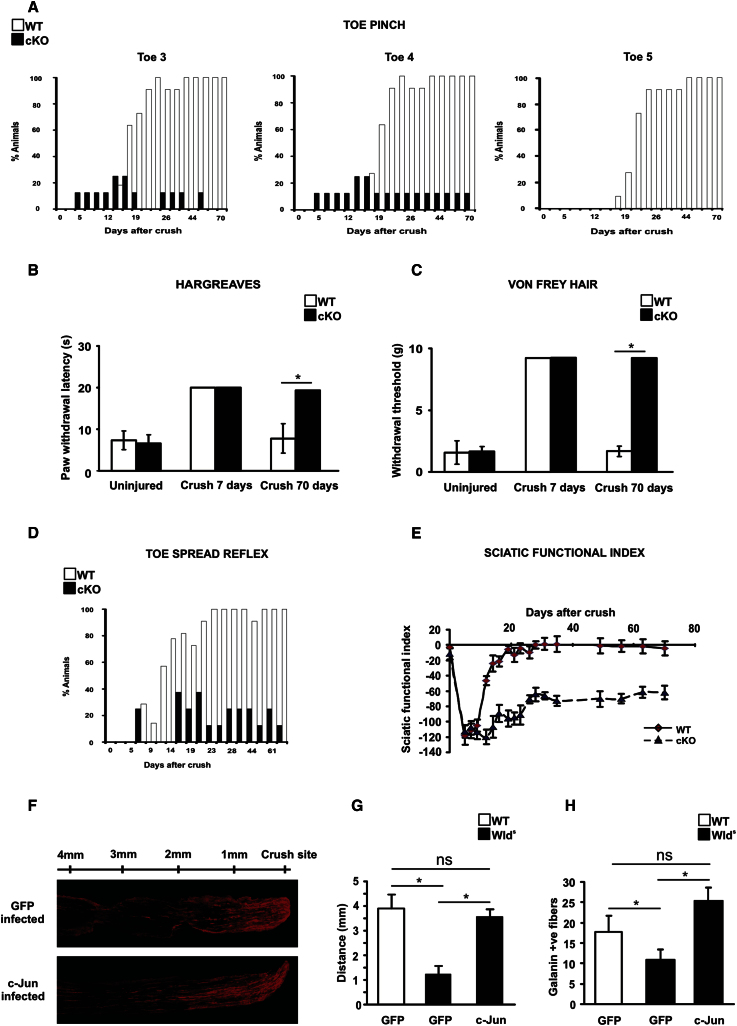
The radical response of peripheral nerves to injury (Wallerian degeneration) is the cornerstone of nerve repair. We show that activation of the transcription factor c-Jun in Schwann cells is a global regulator of Wallerian degeneration. c-Jun governs major aspects of the injury response, determines the expression of trophic factors, adhesion molecules, the formation of regeneration tracks and myelin clearance and controls the distinctive regenerative potential of peripheral nerves. A key function of c-Jun is the activation of a repair program in Schwann cells and the creation of a cell specialized to support regeneration. We show that absence of c-Jun results in the formation of a dysfunctional repair cell, striking failure of functional recovery, and neuronal death. We conclude that a single glial transcription factor is essential for restoration of damaged nerves, acting to control the transdifferentiation of myelin and Remak Schwann cells to dedicated repair cells in damaged tissue.


Figures

Figure 1
Figure 1
c-Jun Controls the Molecular Reprogramming of Schwann Cells in Injured Nerves (A) The number of genes in wild-type (WT) and mutant (cKO) nerves that show significant change in expression levels 7 days post-nerve cut, determined by microarray. The number of up- and downregulated genes is also indicated. Only genes that showed ≥1.5-fold change compared to uninjured nerves were considered. The gene screen data are an average of two independent experiments, each involving nerves pooled from 7–8 animals. (B) Heatmap showing expression of the 172 genes differentially regulated (≥1.5-fold threshold; microarray) in the distal stump of cut WT and c-Jun mutant mice. Comparison of cut WT and cut mutant nerves shows four main types of disregulation in c-Jun mutants (categories 1–4 indicated on the heatmap): (1) enhanced activation, (2) failure of activation, (3) enhanced downregulation, (4) failure of downregulation. (C) Enriched gene ontology (GO) categories for the 172 differentially regulated genes. (D) Subset of the 172 differentially regulated genes chosen for further analysis. This includes the 10 most upregulated and 10 most downregulated genes comparing the distal stump of WT and mutants, and 14 other genes chosen for potential relevance in nerve injury. Genes expressed at higher levels in the distal stump of mutants versus WT are highlighted in red. Genes expressed at lower levels in mutants versus WT are highlighted in blue. FC (cKO/WT) indicates fold change in expression levels in mutant versus WT distal stumps determined by microarray. (E) Genes overexpressed in the mutant: RT-QPCR determination of genes from (D) (red upper panel) showing fold increase in expression in the distal stump of mutants versus WT. Error bars: ± SEM; n = 3–6 pools of 3 animals/pool. (F) Genes underexpressed in mutants: RT-QPCR determination of genes from (D) (blue lower panel) showing fold decrease in expression in the distal stumps of mutants versus WT. Error bars: ± SEM; n = 3–6 pools of 3 animals/pool. See also Figures S1–S4 and Tables S1, S2, S3, and S5.
Figure 2
Figure 2
c-Jun-Dependent Regulation of Schwann Cell Genes and Proteins (A) In purified Schwann cell cultures, c-Jun suppresses myelin genes but activates genes of denervated cells. Graphs show RT-QPCR measurements of six differentially regulated genes in cells from control mice with physiological levels of c-Jun (WT), in cells from mutants lacking c-Jun (cKO), and in mutant cells infected with c-Jun adenovirus to re-express c-Jun (cKO + c-Jun). The y axis shows fold difference in expression levels. Error bars: ± SEM; p < 0.05, n = 4. (B and C) Posttranscriptional control of protein expression by c-Jun. (B) Immunolabeling of Schwann cell cultures (5 days in vitro) from p8 WT and mutant nerves. Three proteins characteristically expressed by denervated Schwann cells are shown. Note suppression of N-cadherin (N-cad) and p75NTR but overexpression of NCAM in mutant cells. (Bar: 50 μm). (C) Western blots of distal stump nerve extracts from WT and mutants (cKO) 7 days after cut show a similar expression pattern. Lower panel: quantitation. Error bars: ± SEM; p < 0.05; ∗∗∗p < 0.001, n = 3. (D and E) Immature and denervated cells differ in gene expression. (D) In situ hybridization using probes against Olig1 (left panel), Shh (middle panel), and GDNF (right panel). mRNAs of these are virtually absent from uninjured newborn and adult sciatic nerve but highly upregulated in distal stumps of adult nerve 3 days after transection. Numerous positive cells display the half moon shape typical of Schwann cells (insets in bottom panels). (Bar: 25μm). (E) Agarose gel electrophoresis of GDNF, Olig1, Shh, and GAPDH products amplified by RT-QPCR from cDNA extracted from WT sciatic nerves of E18 and adult mice and from distal stumps of adult mice 7 days postcut. See also Tables S4 and S5.
Figure 3
Figure 3
c-Jun Controls the Structure of Denervated Schwann Cells In Vivo and in Culture (A) Electron micrographs of distal stumps of WT and c-Jun mutant (cKO) sciatic nerve 28 days after transection (no regeneration). The WT nerve contains classic regeneration tracks (Bands of Bungner; an example is bracketed, showing several Schwann cell processes within a basal lamina tube). These do not form in the mutant, which instead contains irregular and flattened cellular profiles. Bar: 1 μm. (B) The number of cellular profiles per regeneration track is sharply reduced in the mutant. Error bars: ± SEM; p < 0.05, n = 4. (C) Mutant cellular profiles are flatter (lower roundness index). Error bars: ± SEM; p < 0.05, n = 4. (D and E) In vitro, WT Schwann cells from neonatal nerves show typical bi- or tripolar morphology, but mutant cells (cKO) are flat, irrespective of whether they are taken from c-Jun mutants (D) or obtained by infecting c-Junf/f cells with CRE-adenovirus (E). Cells are labeled with β1 integrin antibodies. Bar: 20 μm.
Figure 4
Figure 4
Axonal Injury Results in Extensive Neuron Death in c-Jun Mutants (A and B) Number of unmyelinated axons in L4 dorsal roots (A) and number of small neuronal cell body profiles (B cells) in corresponding DRGs (B), expressed relative to number of B cells in uninjured WT mice. The data show before (0) and at different times after injury. Error bars: ± SEM; p < 0.05, n = 4). (C and D) Number of myelinated axons in L4 dorsal roots (C) and large neuronal cell body profiles (A cells) in corresponding DRGs (D), expressed relative to the number of A cells in uninjured WT mice. The data show before (0) and at different times after injury. Error bars: ± SEM; p < 0.05, n = 4. (E and F) Number of myelinated (E) and unmyelinated (F) axons in tibial nerves (midthigh level) before injury and 10 weeks postcrush in WT and mutant mice. Error bars: ± SEM; p < 0.05, n = 4. Note that axon and neuron numbers are normal in c-Jun mutants before injury, but significantly reduced post nerve crush. See also Figure S2.
Figure 5
Figure 5
Axonal Regeneration Failure in c-Jun Mutants (A) A reduced number of backfilled motoneurons is seen in the ventral horn of c-Jun mutants after True Blue injection into the tibialis anterior muscle 10 weeks post-nerve crush. Bar: 100 μm. (B) Quantification of backfilled neurons 5 and 10 weeks after crush. Abercrombie correction applied. Note reduced number of labeled neurons in the L2–L6 region of the spinal cord. Error bars: ± SEM; ∗∗p < 0.01, n = 4. (C) Regeneration failure in mutants judged by nerve pinch test 4 days post sciatic nerve crush. Error bar: ± SEM; n = 4. (D–H) CGRP+ or galanin+ regenerating axon numbers are reduced in mutants 4 days postcrush. Micrograph (G) shows CGRP labeling of nerve fronts (arrows; bar: 1 mm) in WT and mutant nerves; (H) shows axons 3 mm from the crush site (bar: 100 μm). Regeneration delay is quantified by measuring the distance from crush traveled by the longest axon (D), or by counting how many axons extend 2 or 3 mm from the crush site (E and F). For (D) p < 0.05; ∗∗p < 0.01, n = 4; for (E) and (F) ∗∗p < 0.01, n = 4. (I) In microfluidic chambers, WT Schwann cells and mutant cells with enforced c-Jun expression promote axon growth relative to no cells or mutant cells. Each trace shows axon growth into a side chamber from a central compartment containing neuronal cell bodies. The four types of side chamber are as follows: (no SC) chamber with no Schwann cells; (WT SC) chamber with WT Schwann cells with normal constitutive c-Jun expression; (cKO SC) chamber with cells from c-Jun mutants (no c-Jun), and (cKO SC + c-Jun) chamber with mutant cells infected with c-Jun adenovirus to re-express c-Jun. Bar: 250 μm. (J) Quantification of the number of axons longer than 50 μm growing into the side compartment in all conditions shown in (I). (K) Quantification of the total area covered by axons in the side compartment in all conditions depicted in (I). Error bars: ± SEM; ∗∗p < 0.01, n = 3. See also Figure S2.
Figure 6
Figure 6
Myelin Clearance Is Slow in Injured c-Jun Mutant Nerves (A) Unlike WT nerves, mutant nerves (cKO) are not translucent 4 weeks postcut, indicating lipid retention. Bar: 2 mm. (B) Lipid persistence in mutant nerves shown by quantification of osmium stained area in transverse sections of sciatic nerve 4 weeks post cut (no regeneration). Error bars: ± SEM; p < 0.001, n = 4. (C) Electron micrograph showing lipid droplets in denervated Schwann cells of mutant nerves 4 weeks post cut. Bar: 2 μm. (D) Electron micrograph showing relative preservation of intact myelin sheaths in mutants 3 days postcut, 3 mm from cut site. Right, counts of intact sheaths in tibial nerves of WT and mutants 3 days after cut. Error bars: ± SEM; p < 0.05, n = 5. Bar: 20 μm. (E) Myelin sheath counts in tibial nerves following axotomy in vivo and in nerve segments in vitro, as indicated. Note that sheath preservation in the mutant does not depend on blood-born macrophages. Error bars: ± SEM; p < 0.05, n = 4. (F) Normal myelin breakdown fails in mutant Schwann cells, shown by persistence of myelin debris in mutant cells in phase-contrast micrographs of cultures from p8 WT and mutant nerves and maintained 6 days in vitro. Bar: 50 μm. (G) Immunolabeling of mutant Schwann cells (green; S100 antibodies), bloated with myelin debris (red; MBP antibodies) (arrows indicate two cells). Bar: 50 μm. (H) Counts of cells containing the myelin proteins MPZ and MBP at different times after plating cells from WT and mutant p8 nerves show delay in myelin protein clearance by mutant Schwann cells. 0 = 3 hr after plating. Differences between cKO and WT were significant at all time points. Error bars: ±SEM; p < 0.001, (two way ANOVA), n = 5. (I) Electron micrograph shows the persistence in mutant nerves of large, foamy macrophages (example arrowed). Bar: 5 μm. (J) Lipid droplet counts in macrophages in tibial nerve sections 28 days postcut (no regeneration), show a strong delay in lipid clearance by macrophages in the mutant. Error bars: ±SEM; p < 0.05, n = 4. See also Figures S3 and S5.
Figure 7
Figure 7
Functional Recovery Fails in c-Jun Mutants (A) Percentage of mice responding to pinching of distal parts of toes 3, 4, and 5 after nerve crush. The difference between WT and mutant (cKO) mice was significant from day 21 to day 70, p < 0.01 (two-way ANOVA), n = 5. (B and C) Sensitivity to heat (B), and light touch (C) quantified in mice before and after nerve crush. In animals assayed 7 days after crush, the assay was terminated at 20 s (Hargreaves test) and limited to the use of a hair weight of 8 g (Von Frey test). Mutants show normal sensation when uninjured, but no recovery after injury. Error bars: ±SEM; p < 0.05, n = 4. (D) Percentage of mice showing normal toe spreading reflex (score 0: no toe extension; score 2: full normal extension) after crush. The difference between WT and mutant mice was significant from day 12 to day 70. p < 0.001 (two-way ANOVA), n = 5. (E) Quantification of sensory-motor function in WT and mutants. Note permanent failure of recovery in mutants, although mutant and WT SFIs are similar before and immediately after injury. The difference between WT and mutant mice was significant from day 12 to day 72. Error bars: ± SEM; p < 0.01, n = 4. (F) Increased regeneration in Wlds nerves infected with c-Jun adenovirus compared to GFP control virus, shown by galanin immunolabeling of longitudinal sciatic nerve sections 3 days after crush. (G and H) Regeneration failure in Wlds nerves can be rescued to WT levels by enforced c-Jun expression in Schwann cells. (GFP) nerves infected with GFP control adenovirus; (c-Jun) nerves infected with c-Jun adenovirus; the WT and Wlds genotypes are indicated. Regeneration is measured by the nerve pinch test in (G) and by counting galanin+ axons 3 mm from the crush site (H). Error bars: ± SEM; p < 0.05, ns = not significant), n = 4. See also Figures S2 and S6.

References

    1. Abercrombie M. Estimation of nuclear population from microtome sections. Anat. Rec. 1946;94:239–247. - PubMed
    1. Adameyko I., Lallemend F., Aquino J.B., Pereira J.A., Topilko P., Müller T., Fritz N., Beljajeva A., Mochii M., Liste I. Schwann cell precursors from nerve innervation are a cellular origin of melanocytes in skin. Cell. 2009;139:366–379. - PubMed
    1. Angeloni N.L., Bond C.W., Tang Y., Harrington D.A., Zhang S., Stupp S.I., McKenna K.E., Podlasek C.A. Regeneration of the cavernous nerve by Sonic hedgehog using aligned peptide amphiphile nanofibers. Biomaterials. 2011;32:1091–1101. - PMC - PubMed
    1. Barrette B., Hébert M.A., Filali M., Lafortune K., Vallières N., Gowing G., Julien J.P., Lacroix S. Requirement of myeloid cells for axon regeneration. J. Neurosci. 2008;28:9363–9376. - PMC - PubMed
    1. Chen Z.L., Yu W.M., Strickland S. Peripheral regeneration. Annu. Rev. Neurosci. 2007;30:209–233. - PubMed

Publication types

MeSH terms