Review
. 2012 Nov;8(4):310-26.
doi: 10.2174/157340312803760721.

Triggers and anatomical substrates in the genesis and perpetuation of atrial fibrillation

Affiliations
Review

Triggers and anatomical substrates in the genesis and perpetuation of atrial fibrillation

Damián Sánchez-Quintana et al. Curr Cardiol Rev. 2012 Nov.

Abstract

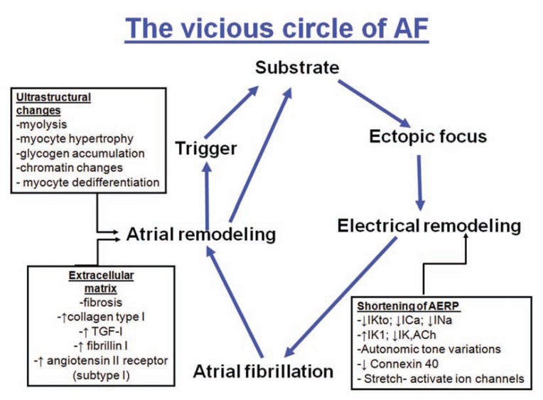
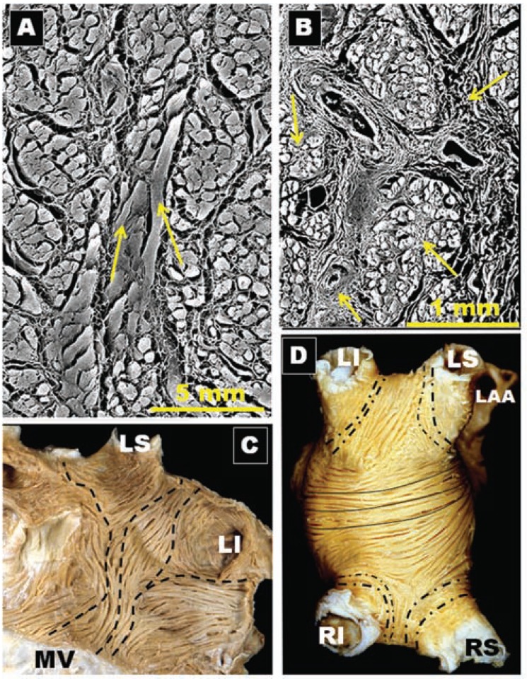
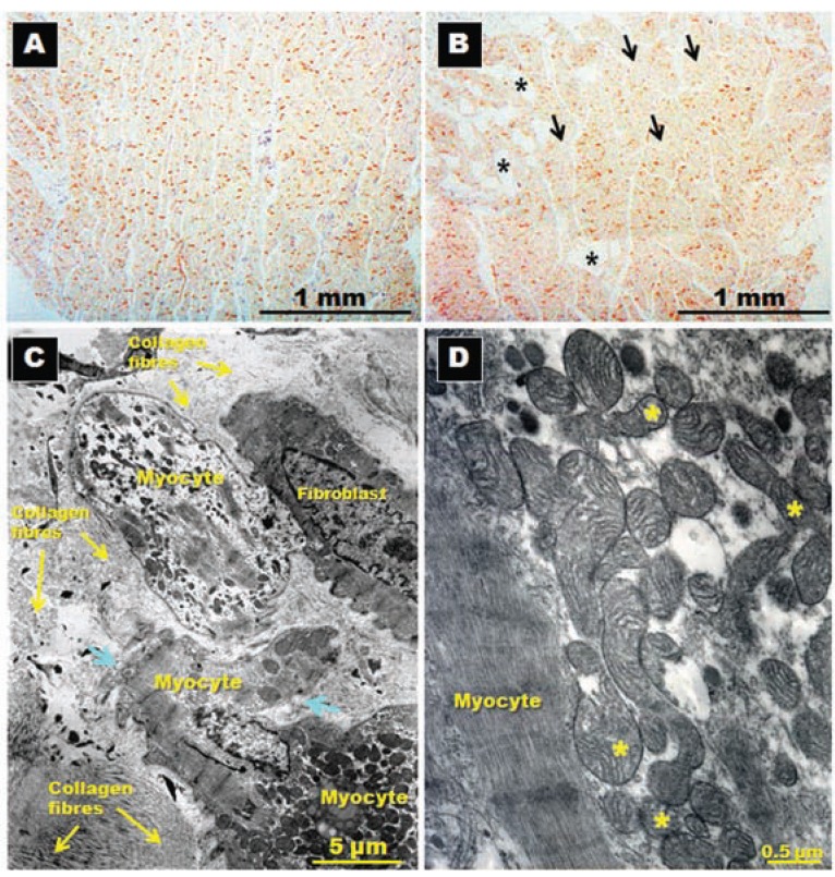
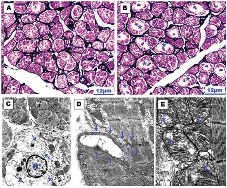
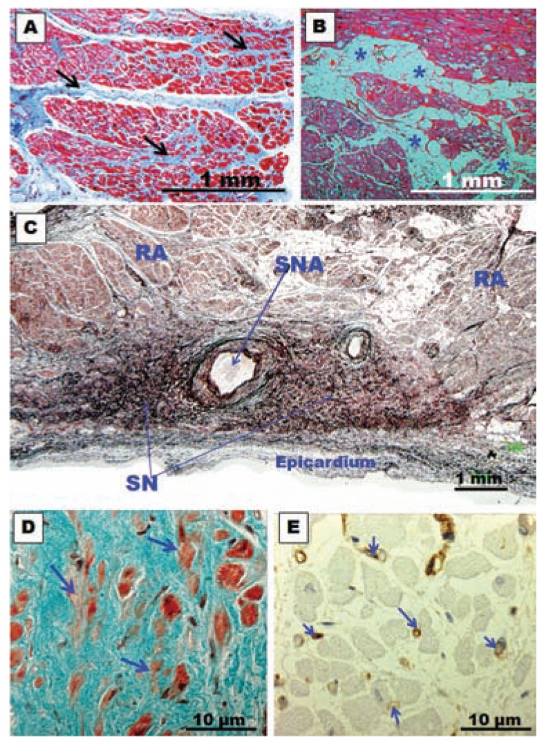
The definition of atrial fibrillation (AF) as a functional electrical disorder does not reflect the significant underlying structural abnormalities. Atrial and Pulmonary Vein (PV) muscle sleeve microstructural remodeling is present, and establishes a vulnerable substrate for AF maintenance. In spite of an incomplete understanding of the anatomo-functional basis for AF, current evidence demonstrates that this arrhythmia usually requires a trigger for initiation and a vulnerable electrophysiological and/or anatomical substrate for maintenance. It is still unclear whether the trigger mechanisms include focal enhanced automaticity, triggered activity and/or micro re-entry from myocardial tissue. Initiation of AF can be favored by both parasympathetic and sympathetic stimulation, which also seem to play a role in maintaining AF. Finally, evolving clinical evidence demonstrates that inflammation is associated with new-onset and recurrent AF through a mechanism that possibly involves cellular degeneration, apoptosis, and subsequent atrial fibrosis.


Figures

Fig. (1)
Fig. (1)
Diagram representing the pathogenesis of the development and maintenance of AF (vicious circle of AF). AERP indicates atrial effective refractory period; IKto, transient outward K+ current; ICa, L-type Ca2+ current; INa, Na+ current; IK1, inward rectifying K+ current; IK, ACh, acetylcholine-regulated K+-current.
Fig. (2)
Fig. (2)
Four hearts specimens with the roof of the left atrium removed and the observer looking towards the mitral orifice. Note the variable pulmonary vein and their venoatrial junctions anatomy between specimens. (A) The arrangement of four pulmonary venous orifices. (B) Left pulmonary veins form a short vestibule (asterisks) or funnel-like common vein before opening left atrium. (C) Three orifices on the right side (arrow). (D) A common vein on the left side. RI, right inferior pulmonary vein; RS, right superior pulmonary vein; LI, left inferior pulmonary vein; LS, left superior pulmonary vein; LCV, left common vein.
Fig. (3)
Fig. (3)
Histological cross-sections with trichrome staining through eight sets of human left pulmonary veins showing the variations in circumferential arrangement of the myocardial sleeves. Note the myocardial sleeve completely surrounding the superior vein and extending beyond 10 mm from the venoatrial junction in a male specimen. The sleeves are thicker in the sectors close to adjacent veins (arrows). Note myocardial strands/ bridges crossing the interpulmonary isthmus or carina between superior and inferior venous sleeves at the level of the venoatrial junction (broken arrow). LI, left inferior pulmonary vein; LS, left superior pulmonary vein;
Fig. (4)
Fig. (4)
Histological cross-sections with trichrome staining through four sets of human right pulmonary veins. In contrast to figure 3, there is no sleeve surrounding the inferior vena from the venoatrial junction to 10 mm. Note the incomplete encirclement of the sleeve at the level of the right superior pulmonary vein (arrows). RI, right inferior pulmonary vein; RS, right superior pulmonary vein.
Fig. (5)
Fig. (5)
Four histological cross-sections with trichrome staining. Note in (A) gap in the myocardial sleeve composed of fibrous tissue (arrow) extending between the adventitia and a media layer of smooth muscle next to endothelium. (B) Interpulmonary myocardial connection at the venoatrial junction (broken arrow). (C) Interpulmonary myocardial connection at the ipsilateral anterior wall of adjacent right pulmonary veins (broken arrow). (D) A small focus of fiber disarray and interstitial fibrosis (asterisks) between the left atrium and left superior pulmonary veins. RI, right inferior pulmonary vein; RS, right superior pulmonary vein; LI, left inferior pulmonary vein; LS, left superior pulmonary vein; LCV, left common vein.
Fig. (6)
Fig. (6)
(A) and (B) Histological cross-sections with trichrome staining in two different specimens. Note the high degree of fibrosis (arrows) of the myocardial sleeve in the left superior pulmonary vein of a specimen with atrial fibrillation. (C) Longitudinal section with van Gieson staining. Note the interstitial fibrosis and scarred peripheral projection of a sleeve (between arrows) acquiring a serpentine shape of a patient with atrial fibrillation. LS, left superior pulmonary vein.
Fig. (7)
Fig. (7)
(A) Low-magnification photomicrograph of superior caval vein (SCV), myocardial sleeves (arrows), right atrial appendage (RAA) and sinus node (SN). RS, right superior pulmonary vein, RCA, right coronary artery (B) Sagittal section of the superior caval vein with trichrome staining. Note the overall architecture of the sinus node (SN) with a radiation located in the superior caval vein (SCV). (C) Histological cross-sections of superior caval vein with trichrome stain showing degenerative changes in a myocardial sleeve in a specimen without history of atrial fibrillation. These changes were more marked in the periphery parts of the myocardial sleeve. (D) The same structural features in C but in a specimen with history of atrial fibrillation.
Fig. (8)
Fig. (8)
(A)Dissection of the left lateral wall of the left atrium to show the myofiber arrangement in the subepicardium of the lateral ridge (LR). Note the vein of Marshall in relationship with LS, left superior; LI, left inferior pulmonary veins. CS, coronary sinus; LAA, left atrial appendage. (B) Sagittal section showing the oblique vein of Marshall and the arrows indicating ganglion and nerve bundles in the vicinity of the vein. (C) Histological section through the left atrioventricular junction shows the coronary sinus (CS) and circumflex artery (LCx) . The CS is surrounded by a sleeve of muscle. There is muscular continuity (arrows) between the sleeve and posterior left atrial wall. MV, mitral valve. (D) Histological cross-sections (Masson trichrome stain) through the coronary sinus (CS) demonstrate the coronary sinus-left atrium muscle connection (asterisks) at the distal end of the coronary sinus. The rest of the myocardial sleeve of the CS is separated from the left atrium wall by epicardial fat. LCx, left circumflex artery; Thv, Thebesian valve; VV, valve of Vieussens.
Fig. (9)
Fig. (9)
(A)Scanning electron micrograph of non-macerated cross section through the body of the terminal crest shows longitudinal fibers (arrows) with intermingling horizontal fibers. (B) Scanning electron micrograph of a cross section through the terminal crest, from a specimen of 70 years old shows a diffuse notable excess of endomysial and perimysial sheaths indicating focal interstitial reactive fibrosis (arrows). (C) Dissection of the subendocardium of the left atrium. The fibers ascend obliquely (broken lines) by the posterior wall of the left atrium and they have an abrupt change of direction at the level of venoatrial junctions surrounding the pulmonary veins. LI, left inferior pulmonary vein; LS, left superior pulmonary vein; MV, mitral valve. (D) The left atrium is everted to show the subendocardial fibers. Note that the fibers pass longitudinally over the roof of the left atrium (continuous lines). Note the abrupt change of direction of circumferential or obliquely fibers (broken lines) around the pulmonary veins. RI, right inferior pulmonary vein; RS, right superior pulmonary vein; LI, left inferior pulmonary vein; LS, left superior pulmonary vein.
Fig. (10)
Fig. (10)
Immunohistochemical staining for N-cadherin and its distribution in posterior left atrial wall (panels A and B) sectioned in longitudinal planes to the long myocyte axis in patients in sinus rhythm (A) and in patients with atrial fibrillation (B). Note the homogenous distribution of N-cadherin in A, whereas some adipocyte cells (asterisks) and patches of myocytes with sparse and reduced levels of N-cadherin (arrows) are present in B. Panels (C) and ((D) Transmission electron micrographs in a specimen with chronic atrial fibrillation for rheumatic mitral valve disease showing in (A) myocytes degeneration surrounded by abundant collagen fibers, myolysis, disruption of basal membrane (arrows) and in (D) abnormal mitochondria with different sizes and alteration of mitochondrial cristae (asterisks).
Fig. (11)
Fig. (11)
(A) and (B)Histological sections of the right atrial appendage with silver-hematoxylin staining. (A) Dog control group in which little myolysis is observed. (B) Dog stimulated group (atrially paced at 400/min for 3 days) in which the myolysis is much more evident around the nucleus of the myocyte (asterisks). (C), (D) and (E) Transmission electron micrographs of the atrial wall observed in a dog subjected to atrial pacing for 3 days. In (C) note the perinuclear myolysis, perinuclear accumulation of glycogen grains (arrows) and nuclear chromatin dispersion. N, nucleus of the myocyte. Magnification x8000. In (D) disruption of an intercalated disc (between arrows). Magnification x10500. In (E) the mitochondria have different sizes and structural alterations with marked dilatation and disruption of mitochondrial cristae (arrows). Magnification x17500.
Fig. (12)
Fig. (12)
(A) and (B)Histological cross-section biopsy samples from the posterior left atrial wall in specimens with chronic atrial fibrillation for rheumatic mitral valve disease. Note in (A) the abundant connective tissue between the myocytes (arrows) and interstitial fibrosis. In (B) note accumulation of fat cells (asterisks) between myocytes. (C) Histological section of the sinus node with methenamine silver staining of a specimen with chronic atrial fibrillation. Note the intense accumulation of connective tissue between nodal cells. SN, sinus node; SNA, sinus node artery; RA, right atrium. (D) Sinus node. Trichrome stain. Note the abundant connective tissue and fewer larger myocardial nodal cells (arrows) in a patient with long-term chronic atrial fibrillation. (E) Immunohistochemical staining for CD31 (vessel walls stained in brown) of the sinus node in a patient with long-term chronic atrial fibrillation. Note fewer and thinner capillaries in the sinus node (arrows).

References

    1. Nattel S. New ideas about atrial fibrillation 50 years on. Nature. 2002;415(10 ):219–26. - PubMed
    1. Haïssaguerre M, Jaïs P, Shah DC, et al. Spontaneous initiation of atrial fibrillation by ectopic beats originating in the pulmonary veins. N Engl J Med. 1998;339(10 ):659–66. - PubMed
    1. Hwang C, Wu TJ, Doshi RN, Peter CT, Chen PS. Vein of Marshall cannulation for the analysis of electrical activity in patients with focal atrial fibrillation. Circulation. 2000;101(13 ):1503–5. - PubMed
    1. Allessie MA, Bonke FI, Schopman FJ. Circus movement in rabbit atrial muscle as a mechanism of tachycardia. III. The leading circle concept: a new model of circus movement in cardiac tissue without the involvement of an anatomical obstacle. Circ Res. 1977;41(1 ):9–18. - PubMed
    1. Wijffels MC, Kirchhof CJ, Dorland R, et al. Atrial fibrillation begets atrial fibrillation. A study in awake chronically instrumented goats. Circulation. 1995;92(7 ):1954–68. - PubMed

Publication types