

Comparative transcriptomic analyses of atopic dermatitis and psoriasis reveal shared neutrophilic inflammation
- PMID: 22920495
- PMCID: PMC3511596
- DOI: 10.1016/j.jaci.2012.06.044
Comparative transcriptomic analyses of atopic dermatitis and psoriasis reveal shared neutrophilic inflammation
Abstract
Background: Atopic dermatitis (AD) and psoriasis are common inflammatory diseases canonically described as involving distinct T(H) polarization and granulocytic infiltration. Acute AD lesions are associated with T(H)2 and eosinophilic inflammation, whereas psoriatic lesions are associated with T(H)1/T(H)17 and neutrophilic inflammation. Despite intensive investigation, these pathways remain incompletely understood in vivo in human subjects.
Objective: Using AD and psoriatic lesional skin as exemplar T(H)2 and T(H)1/T(H)17 diseased tissue, we sought to clarify common and unique molecular and pathophysiologic features in inflamed skin with different types of inflammatory polarization.
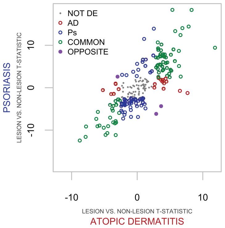
Methods: We conducted gene expression microarray analyses to identify distinct and commonly dysregulated expression in AD (based on Hanifin and Rajka criteria) and psoriatic lesions. We defined gene sets (GSs) as comprising genes encoding cytokines, chemokines, and growth factors that were uniquely or jointly dysregulated in patients with AD and those with psoriasis and calculated aggregate GS expression scores for lesional skin of patients with these dermatoses and healthy control skin.
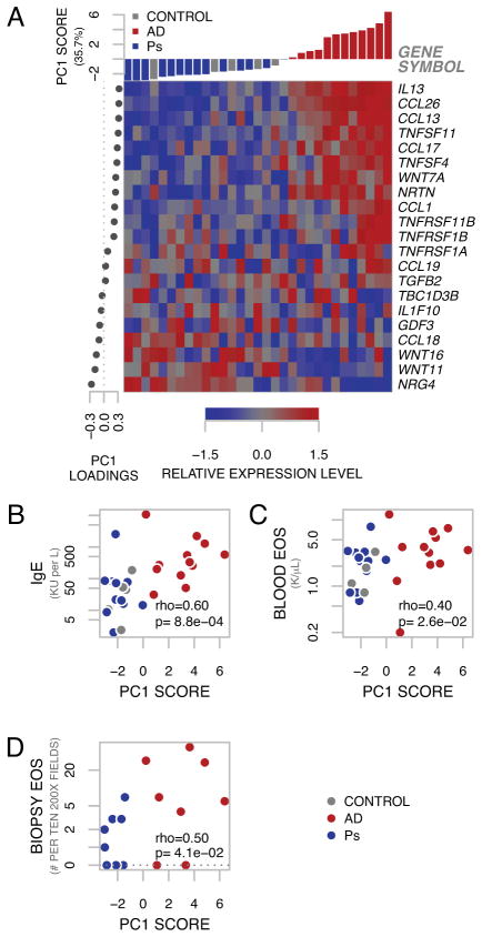
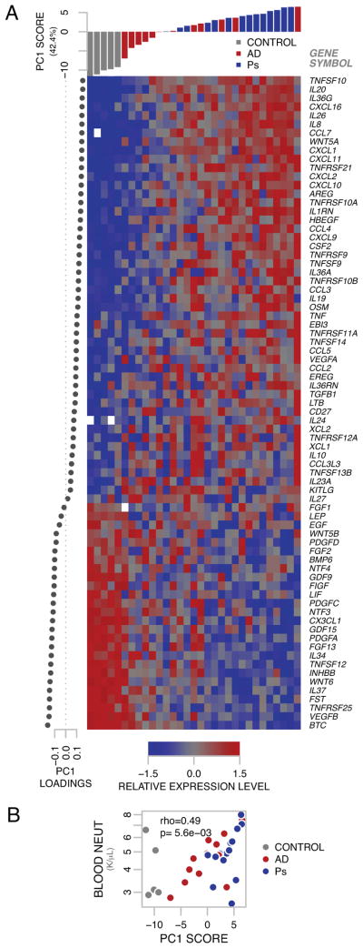
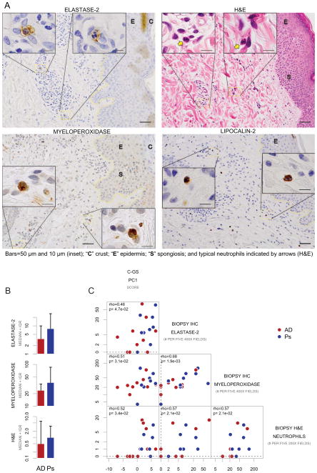
Results: The atopic dermatitis gene set (AD-GS) score correlated with systemic and local measures of allergic inflammation, including serum IgE levels, blood eosinophil counts, and tissue eosinophil counts. Unexpectedly, genes encoding neutrophil chemoattractants among the common GS were highly expressed in AD lesional skin. Hematoxylin and eosin and immunohistochemical analyses showed the numbers of neutrophils in AD lesional skin were comparable with those in psoriatic lesional skin, and both were correlated with the extent of expression of neutrophil chemoattractant genes.
Conclusion: These data are evidence that neutrophilic inflammation is a feature of lesional AD pathology comorbid with allergic inflammation.
Copyright © 2012 American Academy of Allergy, Asthma & Immunology. Published by Mosby, Inc. All rights reserved.
Figures

References
-
- Bieber T. Atopic dermatitis. N Engl J Med. 2008;358:1483–94. - PubMed
-
- Guttman-Yassky E, Nograles KE, Krueger JG. Contrasting pathogenesis of atopic dermatitis and psoriasis--part I: clinical and pathologic concepts. The Journal of allergy and clinical immunology. 2011;127:1110–8. - PubMed
-
- Schon MP, Boehncke WH. Psoriasis. N Engl J Med. 2005;352:1899–912. - PubMed
Publication types
MeSH terms
Substances
Grants and funding
LinkOut - more resources
Full Text Sources
Other Literature Sources
Medical
Molecular Biology Databases
Miscellaneous
