Review
. 2012 Aug 24;37(2):199-207.
doi: 10.1016/j.immuni.2012.08.002.

Regulation of humoral immunity by complement

Affiliations
Review

Regulation of humoral immunity by complement

Michael C Carroll et al. Immunity. .

Abstract

The complement system of innate immunity is important in regulating humoral immunity largely through the complement receptor CR2, which forms a coreceptor on B cells during antigen-induced activation. However, CR2 also retains antigens on follicular dendritic cells (FDCs). Display of antigen on FDCs is critical for clonal selection and affinity maturation of activated B cells. This review will discuss the role of complement in adaptive immunity in general with a focus on the interplay between CR2-associated antigen on B cells with CR2 expressed on FDCs. This latter interaction provides an opportunity for memory B cells to sample antigen over prolonged periods. The cocrystal structure of CR2 with its ligand C3d provides insight into how the complement system regulates access of antigen by B cells with implications for therapeutic manipulations to modulate aberrant B cell responses in the case of autoimmunity.


Figures

Figure 1
Figure 1
Structure-based cartoon depiction of the processes involved in the covalent tagging of antigen with C3b and its subsequent degradation to to the equivalent CR2 ligands iC3b, C3dg and C3d. In native C3, the intramolecular thioester bond of the TED domain is buried at an interface, and is thus denoted in grey. Following proteolytic removal of the C3a activation fragment by C3 convertase, which would be present on the antigen surface, a massive conformational change ensues in the C3b fragment. The activated thioester carbonyl becomes exposed and can react with a hydroxyl group on the antigen surface (grey shaded box), thereby forming a covalent ester linkage. Whereas the CR2 binding surface of the C3d moiety is largely obstructed in C3b, it becomes accessible as a result of the conformational changes accompanying removal of the small C3f peptide of the CUB (for complement C1r and C1s, Uegf and bone morphogenic protein 1) domain by factor I (in concert with cofactor FH) that generates iC3b. An additional cleavage of the CUB domain by FI (this time requiring CR1 (CD35) as the cofactor) releases C3c and leaves C3dg still covalently bound to the antigen. Further proteolytic trimming of C3dg can yield the C3d limit fragment. These cartoons, although greatly simplified to depict only the most relevant domains as individual entities, are based on the crystal structures of native C3 and C3b (Janssen et al., 2006) and EM images of iC3b (Alcorlo et al., 2011; Nishida et al., 2006). The relative binding affinities of the antigen bound C3 split products are indicated.
Figure 2
Figure 2
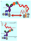
Complement as a bridge linking the innate and adaptive immune systems – the molecular adjuvant role of antigen-linked C3d. A) Co-ligation of the BCR with the CR2;CD19; CD81 complex leads to augmented signaling when naïve B cells first encounter antigen and initiate the process leading to their clonal expansion. The white-boxed area indicates the key binding interaction between CR2(CCP1-2) with a C3d (TED) domain that is covalently bound (yellow triangle) to the antigen recognized by the BCR of this particular B cell. B) CR2 present on FDC may also capture C3d-opsonized antigen and present this antigen to previously primed B cell centrocytes in the germinal centre of the lymph node.
Figure 3
Figure 3
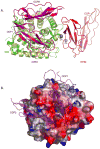
The structure of the CR2 (CCP1-2):C3d complex: A) Comparison of the CR2:C3d binding interfaces in the 2001 (PDB 1GHQ, Szakonyi et al.) and 2011 (PDB 3OED, van den Elsen and Isenman) CR2(CCP1-2):C3d co-crystal structures. Ribbon representations of the essentially identical C3d molecules of the 2001 structure (yellow) and the 2011 structure (green) were superimposed in an orientation where one is looking into the acidic pocket of the C3d molecule. The respective placements of the CR2 (CCP1-2) molecules in the 2001 and 2011 co-crystal structures are denoted by the red and magenta ribbon diagrams, respectively. The grey spheres indicate the respective positions of the two zinc atoms at the interface visualized in the 2001 structure. B) Depiction of the charge complementarity at the interface between the positively-charged side chains of CR2(CCP1-2) that point down towards the acidic pocket on the concave surface of C3d. C3d is shown as a molecular surface representation colored for electrostatic potential; red negative, blue positive, grey neutral. The ribbon rendering of the CR2 domains is semi-transparent. (Adapted from van den Elsen and Isenman, 2011).
Figure 4
Figure 4
Pathways for the recognition of B cell antigen in the lymph node. (1) Immune complexes (ICs), formed by the deposition of complement proteins (in this illustration, C3d) and IgG on the surface of antigen, bind to complement receptor 3 (CR3) on the surface of subcapsular sinus macrophages (MΦ). (2) Naive B cells transport complement-coated ICs from the subcapsular sinus to FDCs. (3) The ICs are transferred in a complement receptor 2 (CR2)-mediated mechanism from the surface of the B cell to the FDC. (4) Cognate B cells capture small antigen directly from the surface of FDCs, associated with CR2 receptors (adapted from (Gonzalez et al., 2011).

Similar articles

Cited by

References

    1. Alcorlo M, Martinez-Barricarte R, Fernandez FJ, Rodriguez-Gallego C, Round A, Vega MC, Harris CL, de Cordoba SR, Llorca O. Unique structure of iC3b resolved at a resolution of 24 A by 3D-electron microscopy. Proc Natl Acad Sci U S A. 2011;108:13236–13240. - PMC - PubMed
    1. Bajenoff M, Germain RN. B cell follicle development remodels the conduit system and allows soluble antigen delivery to follicular dendritic cells. Blood. 2009;114:4989–4997. - PMC - PubMed
    1. Barrault DV, Steward M, Cox VF, Smith RA, Knight AM. Efficient production of complement (C3d)3 fusion proteins using the baculovirus expression vector system. J Immunol Methods. 2005;304:158–173. - PubMed
    1. Boes M, Esau C, Fischer MB, Schmidt T, Carroll M, Chen J. Enhanced B-1 cell development, but impaired IgG antibody responses in mice deficient in secreted IgM. J Immunol. 1998;160:4776–4787. - PubMed
    1. Burman JD, Leung E, Atkins KL, O’Seaghdha MN, Lango L, Bernado P, Bagby S, Svergun DI, Foster TJ, Isenman DE, van den Elsen JM. Interaction of human complement with Sbi, a staphylococcal immunoglobulin-binding protein: indications of a novel mechanism of complement evasion by Staphylococcus aureus. J Biol Chem. 2008;283:17579–17593. - PMC - PubMed

MeSH terms