. 2012 Sep 27;2(3):440-6.
doi: 10.1016/j.celrep.2012.07.015. Epub 2012 Aug 23.

Direct differentiation of human pluripotent stem cells into haploid spermatogenic cells

Affiliations

Direct differentiation of human pluripotent stem cells into haploid spermatogenic cells

Charles A Easley 4th et al. Cell Rep. .

Abstract

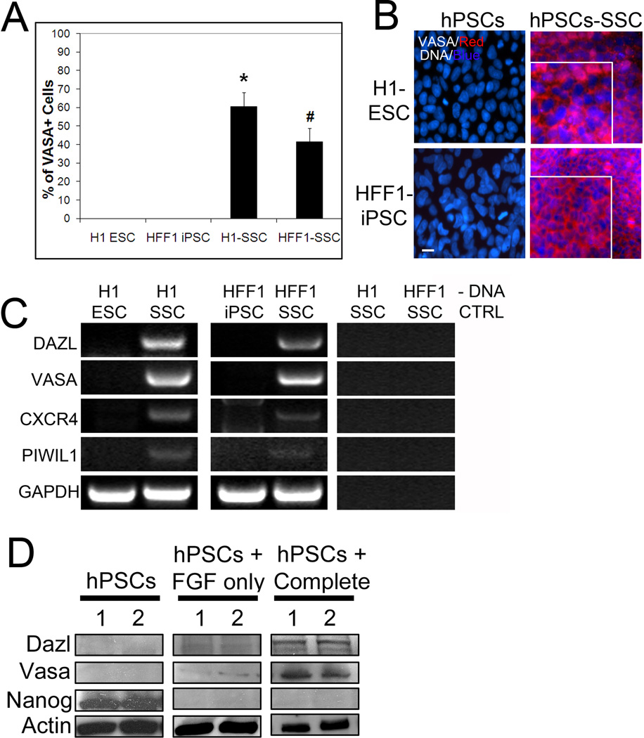
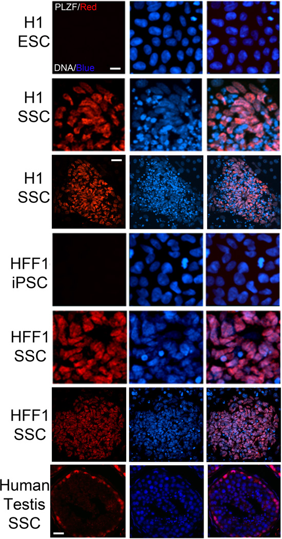
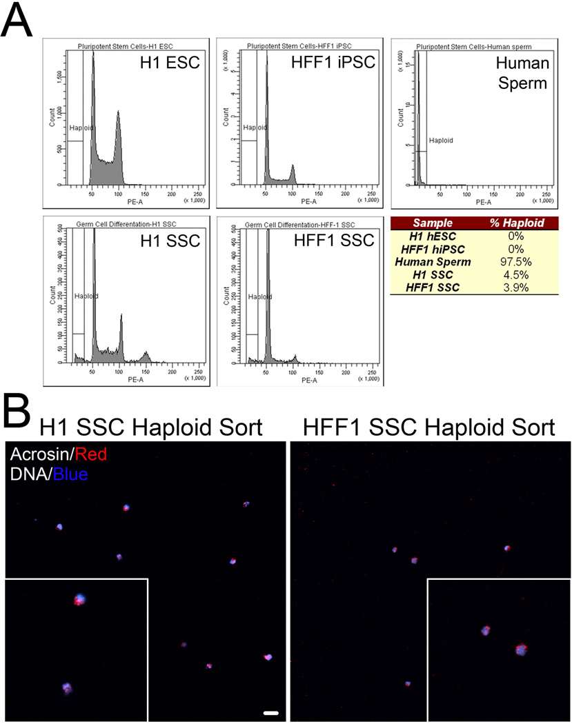
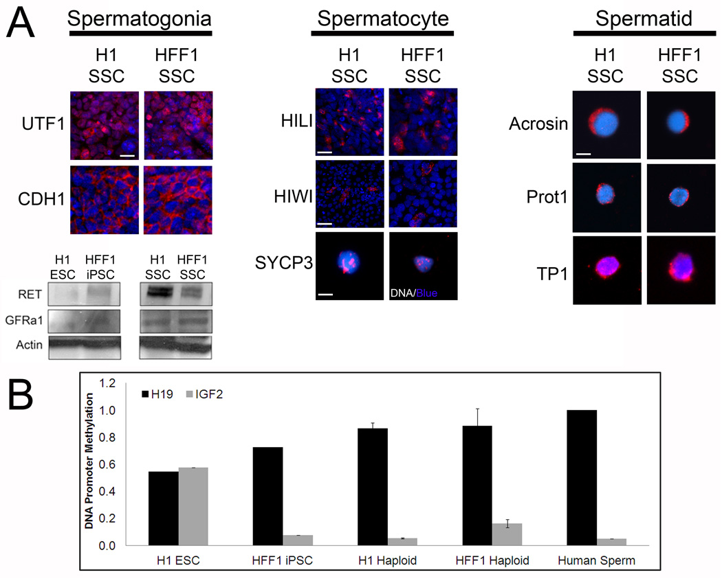
Human embryonic stem cells (hESCs) and induced pluripotent stem cells (hiPSCs) have been shown to differentiate into primordial germ cells (PGCs) but not into spermatogonia, haploid spermatocytes, or spermatids. Here, we show that hESCs and hiPSCs differentiate directly into advanced male germ cell lineages, including postmeiotic, spermatid-like cells, in vitro without genetic manipulation. Furthermore, our procedure mirrors spermatogenesis in vivo by differentiating PSCs into UTF1-, PLZF-, and CDH1-positive spermatogonia-like cells; HIWI- and HILI-positive spermatocyte-like cells; and haploid cells expressing acrosin, transition protein 1, and protamine 1 (proteins that are uniquely found in spermatids and/or sperm). These spermatids show uniparental genomic imprints similar to those of human sperm on two loci: H19 and IGF2. These results demonstrate that male PSCs have the ability to differentiate directly into advanced germ cell lineages and may represent a novel strategy for studying spermatogenesis in vitro.


Figures

Figure 1
Figure 1. Differentiation of hPSCs in spermatogonial stem cell culture yields significant percentages of VASA+ cells
(A) H1 ESCs and HFF-1 iPSCs cultured in mouse SSC conditions for 10 days and then stained for VASA. % VASA expression was quantified in the parent PSC lines and the differentiated lines. Representative graphical analysis from 5 separate trials, > 5000 cells counted for each condition, is shown. * signifies p < 0.01 comparing H1 ESC to H1 SSC. # signifies p < 0.01 comparing HFF1 iPSC to HFF1 SSC. (B) Representative images of PSCs and PSCs differentiated in SSC culture conditions for 10 days and stained for VASA. DNA labeled with Hoechst. Scale, 50 µm. Enlarged insets show typical, perinuclear localization of VASA. (C) Reverse transcription (RT) PCR for germ cell markers DAZL, VASA, CXCR4 and PIWIL1 in PSCs and their differentiated counterparts. GADPH is shown as a loading control. No DNA (-DNA) is also shown as a negative control. (D) Representative western blot analyses showing upregulation of germ cell marker expression and a concomitant loss of the pluripotent marker Nanog in complete SSC culture conditions (with GDNF and FGF). Despite loss of Nanog in FGF only SSC medium (i.e. without GDNF), germ cell markers were not expressed. Actin is a loading control.
Figure 2
Figure 2. Differentiation of hPSCs in SSC conditions results in the expression of the SSC marker PLZF
While the parent PSC lines do not express detectable levels of PLZF, 10 day culture in SSC conditions upregulates PLZF (red) expression in both lines. Hoechst (blue): DNA. Scale, 40 µm. Global view (3rd and 6th rows) of differentiated colonies shows a large portion of cells expressing PLZF. Scale, 100 µm. 7th row panel depicts PLZF staining in human testis sections.
Figure 3
Figure 3. hPSCs differentiated in SSC culture exhibit haploid features
(A) FACS ploidy analysis reveals a small haploid peak in hPSCs cultured in SSC culture conditions for 10 days. This peak corresponds to the haploid peak observed in human sperm. Chart below represents % of haploid cells in undifferentiated and SSC-mediated differentiated hPSCs. Data is representative of five cell sorts with 500,000 cells sorted per experiment. (B) FACS isolated haploid cells from H1 SSC (left) and HFF1 SSC (right) were seeded coverslips and stained with acrosin (red) and Hoechst (DNA, blue). Global view shows several isolated cells with polar acrosin localization. Scale, 50 µm. Insets show zoomed view of acrosin-positive, haploid cells.
Figure 4
Figure 4. Differentiation of hPSCs in SSC culture yields cells that express markers for spermatogonia, spermatocytes and spermatids
(A) Left: 10 days post differentiation cultures of H1 and HFF1 SSCs express pre-meiotic spermatogonial markers UTF1 and CDH1. Scale, 50 µm. Differentiation also induces expression of two membrane receptors: RET and GFRa1. Actin is a loading control. Center: expression of spermatogonia-to-spermatocyte marker HILI, spermatocyte-to-spermatid marker HIWI and meiotic marker SYCP3. Scale for HILI 200 µm, scale for HIWI, 500 µm and scale for SYCP3, 10 µm. Right: expression of post-meiotic spermatid markers Acrosin, Protamine 1 (Prot1) and Transition Protein 1 (TP1). Haploid cells were sorted by FACS and immunostained with antibodies directed at the indicated protein. Scale, 10 µm. (B) H1 ESCs, HFF1 iPSCs, fertile human sperm, and haploid cells obtained by FACS from H1 and HFF1 SSC cultures were examined for methylation on imprinting control regions (ICRs) for H19 (paternally imprinted) and IGF2 (maternally imprinted). Methylation statuses were examined using Qiagen Epitect Methyl II PCR Array. Graph shows average % methylation with error bars.

References

    1. Brinster RL, Avarbock MR. Germline transmission of donor haplotype following spermatogonial transplantation. Proc Natl Acad Sci U S A. 1994;91:11303–11307. - PMC - PubMed
    1. Buaas FW, Kirsh AL, Sharma M, McLean DJ, Morris JL, Griswold MD, de Rooij DG, Braun RE. Plzf is required in adult male germ cells for stem cell self-renewal. Nat Genet. 2004;36:647–652. - PubMed
    1. Bucay N, Yebra M, Cirulli V, Afrikanova I, Kaido T, Hayek A, Montgomery AM. A novel approach for the derivation of putative primordial germ cells and sertoli cells from human embryonic stem cells. Stem Cells. 2009;27:68–77. - PubMed
    1. Carrell DT, Emery BR, Hammoud S. Altered protamine expression and diminished spermatogenesis: what is the link? Hum Reprod Update. 2007;13:313–327. - PubMed
    1. Castrillon DH, Quade BJ, Wang TY, Quigley C, Crum CP. The human VASA gene is specifically expressed in the germ cell lineage. Proc Natl Acad Sci U S A. 2000;97:9585–9590. - PMC - PubMed

Publication types

Substances