. 2013 Jun;7(5):403-11.
doi: 10.1016/j.crohns.2012.08.003. Epub 2012 Aug 23.

Plasminogen activator inhibitor-1 is increased in colonic epithelial cells from patients with colitis-associated cancer

Affiliations

Plasminogen activator inhibitor-1 is increased in colonic epithelial cells from patients with colitis-associated cancer

Earl Gillespie et al. J Crohns Colitis. 2013 Jun.

Abstract

Background: Patients with long-term ulcerative colitis are at risk for developing colorectal cancer.

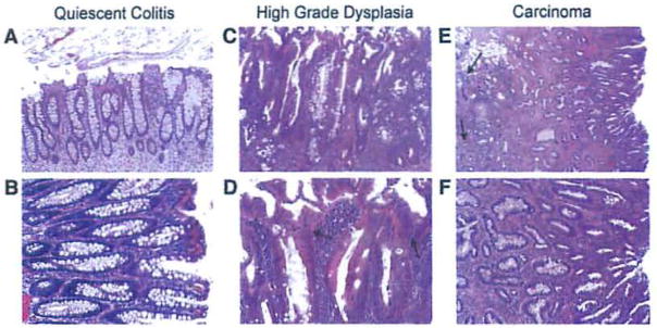
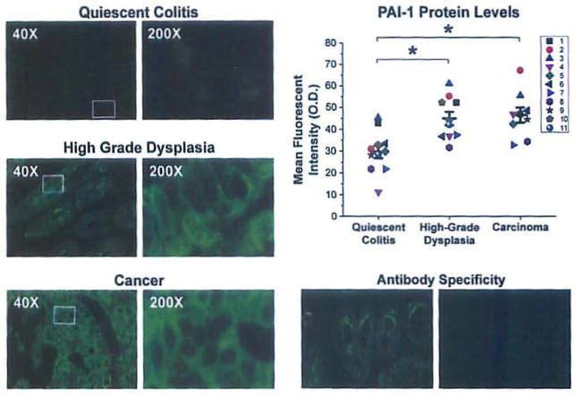
Methods: Archival formalin-fixed paraffin-embedded tissue from ulcerative colitis patients who underwent a colectomy for high-grade dysplasia or carcinoma was examined for changes in expression of plasminogen activator inhibitor-1 (PAI-1) as well as other mediators of inflammation-associated cancer. Epithelia from areas of colons that showed histologic evidence of carcinoma, high-grade dysplasia, and epithelia that were not dysplastic or malignant but did contain evidence of prior inflammation (quiescent colitis) was microdissected using laser capture microscopy. mRNA was extracted from the microdissected tissue and PCR array analysis was performed. To extend our findings, PAI-1 protein levels were determined using immunohistochemistry.

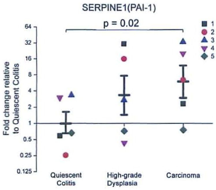
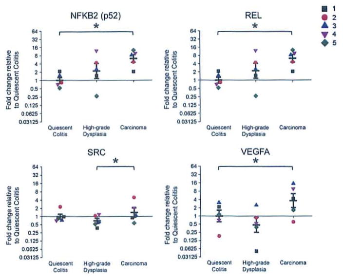
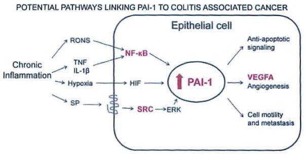
Results: The mRNA expression of PAI-1 is increased 6-fold (p=0.02) when comparing the carcinoma group to the quiescent colitis group; increases were also observed in NFKB2, REL, SRC, and VEGFA. The protein levels of PAI-1 are increased by 50% (p<0.001) in high-grade dysplasia and by 60% (p<0.001) in carcinoma when compared to the quiescent colitis group.

Conclusions: The increase in PAI-1 in high-grade dysplasia and carcinoma suggests a functional role for PAI-1 in malignant transformation in colitis-associated cancer. PAI-1 could also prove a useful diagnostic marker to identify patients at risk for neoplasia and it may be a useful therapeutic target to treat colitis-associated cancer.


Conflict of interest statement

All Authors have no conflict of interest.

Figures

Figure 1
Figure 1
Hematoxylin and eosin stained sections from colitis-associated cancer patients. Bright field photomicrographs from colon section displaying quiescent colitis at (A) 100× and (B) 200× magnification showing branched and elongated crypts typical of reparative processes after inflammation; high-grade dysplasia at (C) 100× and (D)200× magnification; and carcinoma at (E) 40× and (F) 100× magnification. In high-grade dysplasia, note the increased size and disorder of the nuclei (D, indicated by arrows). In carcinoma, note the Invasive pseudo-crypts that extend beyond the mucosal membrane (E, Indicated by arrows).
Figure 2
Figure 2
mRNA expression of SERPINE1 is increased in the transition from quiescent colitis to carcinoma. Each group (n = 5) contains individual observations (each individual is a distinct color/shape) and means ± SEM for groups. Results were calculated using the ΔΔCt method and expressed as a fold change of the quiescent colitis group as described in the RT2 Profile PCR Array Data Analysis (http://www.sabiosciences.com/pcrarraydataanalysis.php).
Figure 3
Figure 3
Protein levels of PAI-1, are increased in the transition from quiescent colitis to high grade dysplasia and carcinoma. Representative images immunofluorescent staining of quiescent epithelium (top left), high-grade dysplasia (middle left) and carcinoma (bottom left) using an antibody targeting PAI-1. Exposure time = 300 msec. Top right, mean fluorescent intensities with individual observations (each individual is a distinct color/shape) and mean ± SEM (n = 11 quiescent colitis, n = 10 high-grade dysplasia, n = 9 carcinoma). Each observation represents the average fluorescent intensity from five randomly selected fields.*p<0.05 compared to quiescent colitis. Bottom right, staining without (left) and with (right) neutralizing peptide displays specificity of the antibody.
Figure 4
Figure 4
mRNA expression of genes that regulate PAI-1 is increased in the transition from non-neoplastic epithelium to carcinoma. Graphs for NKFB2, REL, SRC and VEGFA with individual observations (each patient is a distinct color/shape) with means±SEM. Results were calculated using the ΔΔCt method and expressed as a fold change of the quiescent colitis group as described in the RT2 Profiler™ PCR Array Data Analysis (http://www.sabiosciences.com/pcrarraydataanalysis.php). *p<0.05 difference between groups.
Figure 5
Figure 5
Pathways by which PAI-1 may contribute to colitis-associated cancer. Here we propose a mechanism by which an epithelial cell responds to the stimuli present in chronic inflammation by upregulating PAI-1 and some pro-malignant sequelae of that upregulation. Inflammation produces a variety of signals that can upregulate PAI-1 in colitis-associated cancer; these signals include oxidative stress, inflammatory mediators, hypoxia, and substance P. We have shown that both isoforms of the neurokinin-1 receptor are expressed in colon epithelial cells and that the truncated form is Increased in dysplasia and colitis-associated cancer. The upregulation of PAI-1 has direct consequences for the epithelial cell that promote its transformation to dysplasia or malignancy, including anti-apoptotic signaling and the promotion of anglogenesis and cell motility and metastasis. Additionally, transformed cells may be capable of producing signals that upregulate PAI-1. Genes depicted in bold/red were also identified as increased in this study. Abbreviations: RONS, reactive oxygen and nitrogen species; SP, substance P; HIF, hypoxia inducible factor; NF-κB, nuclear factor kappa B; PAI, plasminogen activator inhibitor; VEGF, vascular endothelial growth factor.

References

    1. O’Connor PM, Lapointe TK, Beck PL, Buret AG. Mechanisms by which inflammation may increase intestinal cancer risk in inflammatory bowel disease. Inflamm Bowel Dis. 2010;16:1411–20. - PubMed
    1. Farraye FA, Odze RD, Eaden J, Itzkowitz SH, McCabe RP, Dassopoulos T, Lewis JD, Ullman TA, James T, III, McLeod R, Burgart LJ, Allen J, Brill JV. AGA medical position statement on the diagnosis and management of colorectal neoplasia in inflammatory bowel disease. Gastroenterology. 2010;138:738–45. - PubMed
    1. Eaden JA, Abrams KR, Mayberry JF. The risk of colorectal cancer in ulcerative colitis: a meta-analysis. Gut. 2001;48:526–35. - PMC - PubMed
    1. Ekbom A, Helmick C, Zack M, Adami HO. Ulcerative colitis and colorectal cancer. A population-based study. N Engl J Med. 1990;323:1228–33. - PubMed
    1. Binder BR, Mihaly J. The plasminogen activator inhibitor “paradox” in cancer. Immunol Lett. 2008;118:116–24. - PubMed

Publication types

MeSH terms

Substances