. 2012 Oct;37(10):404-10.
doi: 10.1016/j.tibs.2012.07.005. Epub 2012 Aug 23.

Flagging and docking: dual roles for N-glycans in protein quality control and cellular proteostasis

Affiliations

Flagging and docking: dual roles for N-glycans in protein quality control and cellular proteostasis

Daniel N Hebert et al. Trends Biochem Sci. 2012 Oct.

Abstract

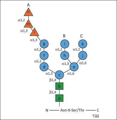
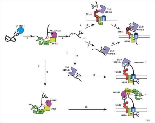
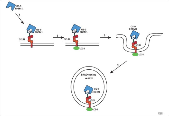
Nascent polypeptides entering the endoplasmic reticulum (ER) are covalently modified with pre-assembled oligosaccharides. The terminal glucose and mannose residues are immediately removed after transfer of the oligosaccharide onto newly synthesized polypeptides. This processing determines whether the polypeptide will be retained in the ER, transported along the secretory pathway, or dislocated across the ER membrane for destruction. New avenues of research and some issues of controversy have recently been opened by the discovery that lectin-oligosaccharide interactions stabilize supramolecular complexes between regulators of ER-associated degradation (ERAD). In this Opinion article, we propose a unified model that depicts carbohydrates acting both as flags signaling the fitness of a maturing protein and as docking sites that regulate the assembly and stability of the ERAD machinery.


Figures

Figure 1
Figure 1
N-Linked glycan structure. The Asn-linked glycan is composed of three glucoses (orange triangles), nine mannoses (blue circles) and two N-acetylglucosamines (green squares). The types of linkage between sugars are denoted and each residue is assigned a letter.
Figure 2
Figure 2
Models of glycoprotein endoplasmic reticulum-associated degradation (ERAD). Various models of the delivery of ERAD substrates to an ER membrane ERAD complex are depicted. Letters A–D designate the different routes for a misfolded substrate. The glycan attached to an ERAD substrate (black sinuous line) is trimmed of a mannose residue by ER mannosidase I (ER Man I, step 1). This is followed by ER-degradation enhancing mannosidase-like protein (EDEM)1 recognition. EDEM1 is present in a complex with ERdj5 and immunoglobulin binding protein (BiP). In the mannose timer model, EDEM1 acts as a mannosidase to trim additional mannose residues (step 2). In route A, ERAD substrates are directly recognized by type I membrane glycoprotein (SEL1L) (step 3). OS-9 and XTP3-B associate with SEL1L to act as ERAD gatekeepers by querying the ERAD substrate for exposed α1,6-linked mannose residues. If the protein possesses the proper glycans, it is passed along the ERAD pathway (step 4). In route B, alternatively, trimmed ERAD substrates are recognized by OS-9 and XTP3-B (step 5) and delivered to the ERAD membrane complex (step 6). Routes C and D depict glycan docking models for OS-9–XTP3-B and EDEM1, respectively. In route C, OS-9 and XTP3-B recognize the misfolded substrates through protein–protein interactions (step 7). This can involve associated co-factors such as GRP94. OS-9 and XTP3-B then deliver the ERAD substrate to the ER membrane ERAD complex by binding to the glycans on the adapter SEL1L (step 8) or on a glycosylated SEL1L associated protein. In route D, the EDEM1 complex associates with ERAD substrates (step 9). EDEM1 employs its mannosidase-like domain to deliver the ERAD substrate to the ER membrane ERAD complex (step 10). The models depicted are not necessarily mutually exclusive. Hybrid models that use multiple pathways are possible. Note that although glycans trimmed to Man7–5 can signal for ERAD in metazoans, the Man6 composition is displayed for simplicity.
Figure 3
Figure 3
Endoplasmic reticulum-associated degradation (ERAD) tuning model. The receptor-mediated removal of ER-degradation enhancing mannosidase-like protein (EDEM1) and OS-9 from the ER lumen is shown. In the absence of misfolded proteins, type I membrane glycoprotein (SEL1L) is disengaged from the HRD1 dislocation machinery. The penta-glycosylated ectodomain of SEL1L associates with EDEM1 and OS-9 (step 1). The cytosolic tail of SEL1L or a SEL1L-associated protein binds the cytosolic ubiquitin-like protein LC3-I. The complex is released from the ER in vesicles (steps 3 and 4) that may eventually fuse with an ill-defined degradative compartment. ERAD tuning vesicles are co-opted by coronaviruses as replication and transcription platform vesicles.

Similar articles

Cited by

References

    1. Zielinska D.F. Precision mapping of an in vivo N-glycoproteome reveals rigid topological and sequence constraints. Cell. 2010;141:897–907. - PubMed
    1. Helenius A., Aebi M. Roles of N-linked glycans in the endoplasmic reticulum. Annu. Rev. Biochem. 2004;73:1019–1049. - PubMed
    1. Lederkremer G.Z. Glycoprotein folding, quality control and ER-associated degradation. Curr. Opin. Struct. Biol. 2009;19:515–523. - PubMed
    1. Hebert D.N. ERAD substrates: which way out? Semin. Cell Dev. Biol. 2010;21:526–532. - PubMed
    1. Aebi M. N-Glycan structures: recognition and processing in the ER. Trends Biochem. Sci. 2010;35:74–82. - PubMed

Publication types

LinkOut - more resources