

Determination of ureter stent appearance on dual-energy computed tomography scan
- PMID: 22921702
- PMCID: PMC3939716
- DOI: 10.1016/j.urology.2012.07.005
Determination of ureter stent appearance on dual-energy computed tomography scan
Abstract
Objective: To examine the dual-energy computed tomography (DECT) properties of 7 commonly used ureteral stents to optimize stent selection for calculi monitored using DECT. The use of DECT to evaluate renal and ureteral calculi has recently increased.
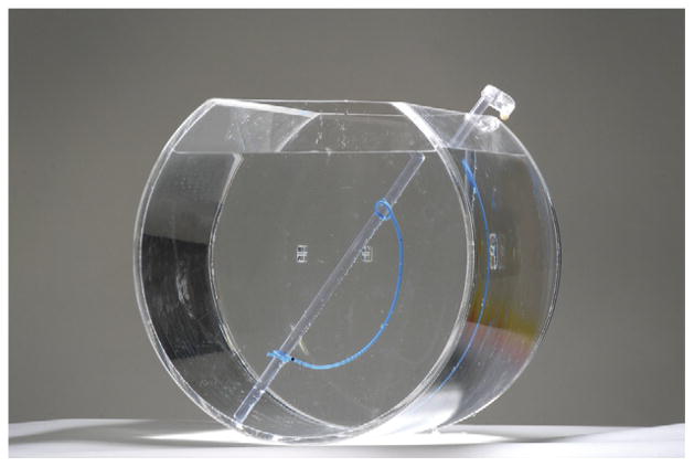
Methods: Seven stents were individually placed in a fish bowl phantom and imaged using a Siemens Somatom Definition Flash CT scanner. DECT peak tube potentials of 80 and 140 kVp and 100 and 140 kVp were used, reflecting our current dual-energy protocols. These were compared to 31 in vivo stents of known composition. The data were reconstructed on a multimodality WorkPlace (Siemens) using CT syngo Post-Processing Suite software.
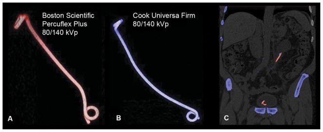
Results: The average patient age was 64 years (range 27-90). The average body mass index was 31.9 kg/m(2) (range 24-51.6). Of the 27 patients, 4 had uric acid stones and 22 had calcium-based stones; 1 patient had undergone renal transplantation. No difference was seen in the dual-energy characterization of stents from the same manufacturer. All imaged Cook and Bard stents had a dual-energy characterization that approached that of calcium stones (blue). All Boston Scientific and Gyrus ACMI stents had a dual-energy characterization resembling that of uric acid stones (red).
Conclusion: The present study evaluated the stent appearance on DECT for various stent manufacturers. This information will aid in the optimal stent selection for patients undergoing treatment of renal calculi and followed up with DECT.
Copyright © 2012 Elsevier Inc. All rights reserved.
Figures
References
-
- Boll DT, Patil NA, Paulson EK, et al. Renal stone assessment with dual-energy multidetector CT and advanced postprocessing techniques: improved characterization of renal stone composition—pilot study. Radiology. 2009;250:813–820. - PubMed
-
- Eiber M, Holzapfel K, Frimberger M, et al. Targeted dual-energy single-source CT for characterisation of urinary calculi: experimental and clinical experience. Eur Radiol. 2012;22:251–258. - PubMed
-
- Eliahou R, Hidas G, Duvdevani M, et al. Determination of renal stone composition with dual-energy computer tomography: an emerging application. Semin Ultrasound CT MR. 2010;31:315–320. - PubMed
-
- Graser A, Johnson TRC, Bader M, et al. Dual energy CT characterization of urinary calculi: initial in vitro and clinical experience. Invest Radiol. 2008;43:112–119. - PubMed
-
- Matlaga BR, Kawamoto S, Fishman E. Dual source computer tomography: a novel technique to determine stone composition. J Urol. 2008;5:1164–1168. - PubMed
