. 2012 Oct 10;20(10):1692-703.
doi: 10.1016/j.str.2012.07.015. Epub 2012 Aug 23.

Enterohaemorrhagic Escherichia coli exploits a tryptophan switch to hijack host f-actin assembly

Affiliations

Enterohaemorrhagic Escherichia coli exploits a tryptophan switch to hijack host f-actin assembly

Olli Aitio et al. Structure. .

Abstract

Intrinsically disordered protein (IDP)-mediated interactions are often characterized by low affinity but high specificity. These traits are essential in signaling and regulation that require reversibility. Enterohaemorrhagic Escherichia coli (EHEC) exploit this situation by commandeering host cytoskeletal signaling to stimulate actin assembly beneath bound bacteria, generating "pedestals" that promote intestinal colonization. EHEC translocates two proteins, EspF(U) and Tir, which form a complex with the host protein IRTKS. The interaction of this complex with N-WASP triggers localized actin polymerization. We show that EspF(U) is an IDP that contains a transiently α-helical N-terminus and dynamic C-terminus. Our structure shows that single EspF(U) repeat forms a high-affinity trimolecular complex with N-WASP and IRTKS. We demonstrate that bacterial and cellular ligands interact with IRTKS SH3 in a similar fashion, but the bacterial protein has evolved to outcompete cellular targets by utilizing a tryptophan switch that offers superior binding affinity enabling EHEC-induced pedestal formation.


Figures

Figure 1
Figure 1. Structural characterization of free EspFU
A) Amino acid sequence of EspFU fifth repeat (R475) along with N-WASP GBD and IRTKS/IRSp53 SH3 binding epitopes. B) Structural disorder prediction for R475 based on IUPred and Disprot algorithms. C) 15N-1H correlation (HSQC) spectrum of 15N,13C labeled EspFU R475, recorded at 800 MHz 1H frequency. Narrow range of 1H chemical shifts is a signature of disordered nature of EspFU R475. D) Analysis of 13Cα and secondary chemical shifts in unbound EspFU R475. Deviations from residue specific random coil chemical shifts are shown, which take into account the nearest neighbor effects and temperature (Kjaergaard & Poulsen, 2011). E) Values of reduced spectral density functions at three frequencies 0.87ωH, ωN and 0 against the primary sequence of EspFU R475. Transiently populated α-helix as well as XPxXP motifs are shown above histograms. See also Figures S1 and S2.
Figure 2
Figure 2. Structure and dynamics ternary complex
Structure of trimolecular complex between N-WASP GBD, EspFU R475 and IRTKS SH3. A) Ribbon presentation of the lowest energy conformation of the ternary complex. N-WASP GBD orange, EspFU R475 green and IRTKS SH3 magenta. B) Superimposition of 20 lowest energy conformers of N-WASP GBD 212–270: EspFU 502–521(left) and IRTKS SH3 343–400: EspFU 527–540 (right). Same coloring as in A). C) Superimposition of GBD domains from N-WASP:EspFU (red) and the WASP:EspFU complexes (Cheng et al., 2008) (blue) for residues 216–262. M238, C239 from N-WASP GBD and the corresponding R35 and A36 from WASP are shown in stick presentation. D) Chemical shift perturbations observed 1H-15N HSQC spectra between unbound EspFU R475 (blue contours), in complex with IRTKS SH3 (red contours) and in complex with N-WASP GBD and IRTKS SH3 (green contours). E) Comparison of steady state heteronuclear {1H}-15N NOEs for unbound EspFU R475 (blue bars) and associated to trimolecular complex with N-WASP GBD and IRTKS SH3 (red bars). F) Color coding of observed {1H}-15N NOEs in the complex on the structure of EspFU R475 reflects the increased rigidity on ps-ns timescales for N-WASP binding residues 3–21 and IRTKS SH3 binding epitope H26-V40 (blue coloring). Sustained flexibility on ps-ns timescales (colored green and yellow) is observed for the linker residues 22–24 that connect GBD and SH3 binding domains. The C-terminal end of EspFU R475 remains highly disordered also in the complex. See also Figure S3.
Figure 3
Figure 3. Chemical shift perturbation mapping of IRTKS SH3 upon addition of peptides from EspFU and Eps8
Full assignment of resonances is given for IRTKS SH3:EspFU R475 complex (upper left panel). Selected boxed resonances with assignments capture the embedded binding affinity and show that all the peptides bind to the same binding site on IRTKS SH3. The assignments corresponds to the saturated state with IRTKS SH3:peptide shown in blue. Saturated state for EspFU R475 and Eps8A33W correspond to SH3:peptide molar ratio 1:1 and for EspFUW33A and Eps8 to SH3:peptide molar ratio 1:3.25. Free IRTKS resonances are shown in red and other colors correspond to intermediate states between free and saturated states.
Figure 4
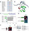
Figure 4. Structural and functional role of the W-switch
A) Alignment of tandem PxxP containing ligands. Delphilin is a protein that we predicted as a potential novel IRTKS ligand, but this has not been experimentally tested. B) EspFU W33 establishes a T-shaped edge-to-face arrangement with IRTKS W378. C) Schematic showing a single repeat of EspFUC. The N-WASP binding helix “H” (Cheng et al., 2008) and IRTKS binding “P” (Weiss et al., 2009; Vingadassolom et al., 2009) domains are indicated. Asterisk indicates the site of the W33A mutation. D) Plasmids encoding the LexA DNA binding domain-IRTKSSH3 and the indicated GAL4 AD-EspFU fusions were co-transformed into a yeast two-hybrid reporter strain L40. “PW33A” indicates the mutant with the alanine substitution of residue W33. β-galactosidase activity was assessed as an average of three co-transformants in Miller Units (MU) with error bars indicating the standard deviation. Results are representative of at least three experiments. D) FLCs expressing myc-tagged GFP-EspFU fusions were infected with EPEC KC12, which requires ectopic expression of EspFU for pedestal formation. Red asterisks (and corresponding red stripes in schematic) indicate W33A mutations. Monolayers were stained with DAPI (blue), anti-myc antibody (green), and Alexa568-phalloidin (red). E) FLCs expressing HA-tagged Tir-EspFU fusion protein carrying W33A mutations were infected with intimin-expressing E. coli K12. Monolayers were stained with DAPI (blue), anti-HA antibody (green), and Alexa568-phalloidin (red). F) An engineered tryptophan mutation in Eps8 dramatically enforces its intracellular association with IRTKS. Human 293T cells were transfected with an expression vector for IRTKS tagged with a biotin acceptor domain together with a vector for a Myc-tagged wild-type Eps8 (wt) or a mutated derivative (mut) containing an EspFU-like tryptophan-containing linker between the PxxP motifs in the IRTKS SH3 domain binding region. IRTKS from lysates of these cells was precipitated using streptavidin-coated beads. Proteins precipitated from these lysates with streptavidin-coated beads were examined by Western blotting using anti-Myc antibodies and labeled streptavidin to detect Eps8 and IRTKS proteins, respectively. Part of the total lysates was similarly analyzed for Eps8 and IRTKS expression without prior affinity selection, as indicated.

Comment in

Similar articles

Cited by

References

    1. Aitio O, Hellman M, Kazlauskas A, Vingadassalom DF, Leong JM, Saksela K, Permi P. Recognition of tandem PxxP motifs as a unique Src homology 3-binding mode triggers pathogen-driven actin assembly. Proc Natl Acad Sci USA. 2010;107:21743–21748. - PMC - PubMed
    1. Babu MM, van der Lee R, de Groot NS, Gsponer J. Intrinsically disordered proteins: regulation and disease. Curr Opin Struct Biol. 2011;21:432–440. - PubMed
    1. Bissantz C, Kuhn B, Stahl M. A medicinal chemist’s guide to molecular interactions. J Med Chem. 2010;53:5061–5084. - PMC - PubMed
    1. Campellone KG, Robbins D, Leong JM. EspFU is a translocated EHEC effector that interacts with Tir and N-WASP and promotes Nck-independent actin assembly. Dev Cell. 2004;7:217–228. - PubMed
    1. Campellone KG, Leong JM. Nck-independent actin assembly is mediated by two phosphorylated tyrosines within enteropathogenic Escherichia coli Tir. Mol Microbiol. 2005;56:416–432. - PubMed

Publication types

MeSH terms

Associated data