. 2012 Oct 10;20(10):1726-36.
doi: 10.1016/j.str.2012.07.014. Epub 2012 Aug 23.

Structural basis for the versatile interactions of Smad7 with regulator WW domains in TGF-β Pathways

Affiliations

Structural basis for the versatile interactions of Smad7 with regulator WW domains in TGF-β Pathways

Eric Aragón et al. Structure. .

Abstract

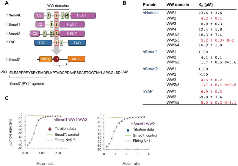
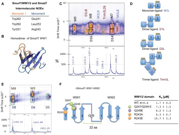
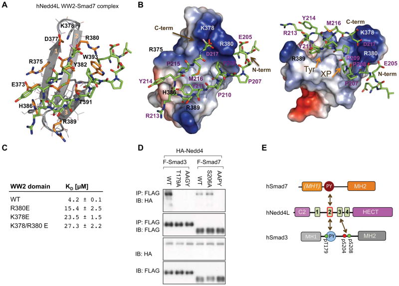
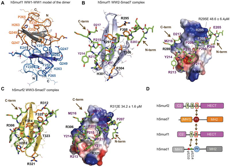
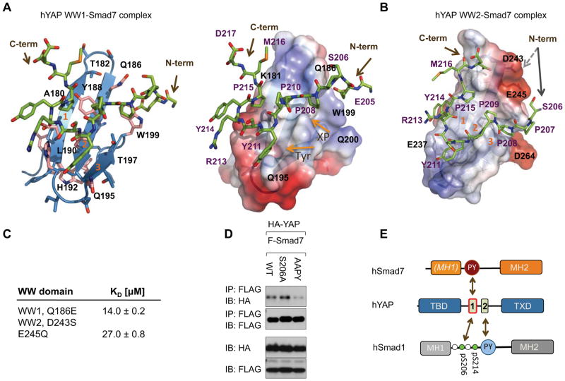
Transforming growth factor (TGF)-β and BMP signaling is mediated by Smads 1-5 (R-Smads and Co-Smads) and inhibited by Smad7, a major hub of regulation of TGF-β and BMP receptors by negative feedback and antagonistic signals. The transcription coactivator YAP and the E3 ubiquitin ligases Smurf1/2 and Nedd4L target R-Smads for activation or degradation, respectively. Pairs of WW domain in these regulators bind PY motifs and adjacent CDK/MAPK and GSK3 phosphorylation sites in R-Smads in a selective and regulated manner. In contrast, here we show that Smad7 binds YAP, Smurf1, Smurf2, and Nedd4L constitutively, the binding involving a PY motif in Smad7 and no phosphorylation. We also provide a structural basis for how regulators that use WW domain pairs for selective interactions with R-Smads, resort to one single versatile WW domain for binding Smad7 to centralize regulation in the TGF-β and BMP pathways.


Figures

Figure 1
Figure 1. Domain composition of the human Nedd4L, Smurf1, Smurf2, YAP and Smad7 proteins and binding affinities, determined by Isothermal Titration Calorimetry
(A) The three-ubiquitin ligases contain the characteristic C2 domain, a central region with a variable number of WW domains and the catalytic HECT domain, each domain represented as a rectangle. The human Yes Associated Protein (YAP) contains a TEA binding domain, two WW domains and a transactivator domain (TXD). Smad7 contains a canonical MH2 group and a divergent MH1. To highlight this divergence we have labeled the MH1 domain using italics and in brackets. The detailed sequence of the Smad7 linker is shown (residues 203–248). The synthesized PY peptide is underlined. The WW domains that mediate the interaction with the PY motif of Smad7 are indicated with an arrow. (B) ITC affinity values for the recombinant fragments of Nedd4L, Smurf1, Smurf2 and YAP and the Smad7 peptide. Binding experiments have been performed at least three times, using two protein expression batches, and different buffers and temperatures. Values hereby presented were obtained at 15 °C. (C) ITC curves for the Smurf1 WW1-WW2 and the independent WW2 fragments in the presence of the Smad7 peptide. The data represented here correspond to values acquired at 15 °C in Tris buffer. The affinity values were 1.7 ± 0.3 μM and 4.1 ± 0.6 μM with stoichiometries of 0.7 and 1 respectively.
Figure 2
Figure 2. Smurf1WW1-WW2 and Smurf2WW2-WW3 form different species in the presence of Smad7 peptide
(A) Assigned NOEs defining the hSmurf1WW1-WW1 dimer interface. Schematic representations of the possible species present in the complexes of Smurf1 WW1-WW2 pair with Smad7. (B) Schematic representation of the Smurf1 WW1 dimer. Secondary structure elements are shown in gray and in blue shades and numbered. The model has been generated using the structure of the published Smurf1 WW1 domain (Aragón et al., 2011) pdb entry: 2laz and the set of assigned inter-molecular NOEs. (C) An expansion of the Ion Mobility-Mass Spectrometry data obtained for the complexes of Smurf2WW2-WW3 with the Smad7 peptide displaying nine characterized species (the full spectrum is shown as Figure S1). Each ion was assigned to a given species based on its characteristic mobility. Abbreviations used are ML (Monomer with one ligand), D1L and D2L (Dimer with one or two ligands respectively), Trim1L, Trim2L and Trim3L (Trimers with one, two or three ligands). Numbers following the specie’s name reflect the protonation state. We have unambiguously detected dimers with one or with two bound ligands and trimers in both Smurf1 and Smurf2 complexes. (D) Schematic representation of the species identified by Ion Mobility-Mass Spectrometry (IM-MS) for the complexes of Smurf1 WW1-WW2 and Smurf2WW2-WW3 pairs with the Smad7 PY site. The WW domains are represented as blue rectangles (labeled in black, white and violet to represent monomer, dimer and trimers respectively). The Smad7 peptide is represented as a green thick-line on top of the WW2 domain. Contacts involving two WW1 domains or between the WW1 domain of one molecule with the linker connecting the WW pair of a second molecule are based on experimental NOEs. (E) A region of the Ion Mobility-Mass Spectrometry data obtained for the Smurf1 WW1 dimer, (the full spectrum is shown as Figure S1). As in (C), each ion was assigned to a given species based on its characteristic drift-time. (F) Schematic representation of the pair of WW domains and the linker present in Smurf1. The mutated positions used in the ITC binding experiments and the three strands of each WW domain are labeled. Next to it, the list of the measured affinity values.
Figure 3
Figure 3. Structure of the Nedd4L WW2 domain bound to the Smad7 linker
(A) Detailed view of the refined structure of the Nedd4L WW2 domain (364–403) in complex with the Smad7 synthesized PY fragment (203–217). The elements of secondary structure (graphite) are numbered and key residues of the Nedd4L WW2 domain that participate in the interaction with the peptide are labeled and highlighted in orange. (B) Electrostatic potential surface of the Nedd4L WW2 domain with the bound Smad7 peptide (stick representation) colored in green. The lowest energy structure displayed in (A) and (B) panels and the family of 25-calculated structures are shown as Figure S2 A and B. Positively charged sites are colored in blue and negatively charged sites in red. Key residues in Smad7 and Nedd4L are indicated (violet and black, respectively). The N- and C-term sites of the Smad7 peptide are indicated with an arrow. (C) Two residues that recognize the N-term part of Smad7 were both independently and jointly mutated to glutamic acid, and the effect of the change in the interaction affinity was determined by ITC. The binding affinity decreased by approximately 7-fold with respect to the wild-type Nedd4L WW2 domain. (D) Human HEK293T cells transduced with vectors encoding the indicated epitope-tagged proteins were subjected to immunoprecipitation (IP) with anti-Flag antibodies and western immunoblotting with antibodies against the indicated tags (upper panels). Aliquots of cell lysates were directly subjected to immunoblotting as loading controls (bottom panels). (E) Schematic representation of the different binding modes displayed by Nedd4L with Smad3 and Smad7. Nedd4L binds preferentially to the PY site of Smad7 using its WW2 domain (this work) while binding to Smad3 region including the di-phosphorylated site and pT[PY] motif requires the WW2-WW3 pair (Aragón et al., 2011).
Figure 4
Figure 4. Structure of the Smurf1 WW2 and Smurf2WW3 domains bound to the Smad7 linker
(A) A detailed view of the binding interface of the dimer between two Smurf1 WW1 domains (residues 233–270). The orientation is rotated by 90 degrees with respect to the schematic representation shown in Figure 2B. The unambiguously assigned NOEs that defined the dimer interface are represented with dotted lines. Monomers are colored in orange-gray (top) and cobalt blue (bottom), with key residues highlighted. (B) Refined structure of the Smurf1 WW2 domain (residues 277–314, light-blue) bound to the Smad7 peptide (203–217, green). Next to it is the charge distribution on the surface of the Smurf1 WW2 domain in complex with the peptide. The structure with the lowest energy was selected for both representations. The family of 25-calculated structures is shown as Figure S3A. N- and C-termini of Smad7 peptide are represented with brown arrows. Key residues that participate in the complex interaction are labeled in purple (Smad7) and in black (Smurf1). The gray line indicates the position of the R295E mutation, which decreases the binding affinity of the complex by approximately 8-fold. (C) Same representations as above for the Smurf2 WW3 domain (297–333, gold) bound to the Smad7 peptide (green). The R312E mutation, which decreases the binding affinity of the complex by approximately 10-fold, is indicated with a gray line. The family of 25-calculated structures is shown as Figure S3B. (D) Schematic representation of the different binding modes observed for Smurf1 and Smurf2 WW domains with respect to R- and I-Smad linkers. Both Smurf proteins use a unique domain to interact with Smad7 (this work) while Smurf1 uses the WW1-WW2 pair to interact with a di-phosphorylated Smad1 PY containing site (Alarcón et al., 2009; Aragón et al., 2011).
Figure 5
Figure 5. Structure of the YAP WW1 and WW2 domains bound to the Smad7 linker
(A) Detailed view of the interaction of the YAP WW1 domain (residues 163–206, blue) with the Smad7 [PY] peptide (green) and next to it, the charge distribution of the domain in the complex, shown as a semitransparent surface representation. The structure with the lowest energy was selected for both representations. The family of 25-calculated structures is shown as Figure S4A, B. Critical residues involved in the interaction are labeled in purple (Smad7) and in black (YAP WW1). Both N- and C-termini of Smad7 peptide) are represented with arrows. (B) A semitransparent surface representation showing the charge distribution of the YAP WW2 domain (residues 227–266, deep blue) bound to the Smad7 peptide represented as sticks (green). The family of 25-calculated structures is shown as Figure S4B. The peptide’s N-terminus (Ser206) is shifted from the loop1 with respect to the orientation in the complex with WW1. The different positions of the peptide in both complexes are shown with two arrows, with a straight gray line (WW2 complex) and with a dotted gray line (the position in the WW1 complex). (C) Effect of point mutations in the YAP WW1 and WW2 domains. The Q186E change in the WW1 domain reduces the affinity by two-fold, while a double change introduced in the WW2 loop -to mimic the sequence of the first WW domain- improves the affinity by a factor of two. Mutated residues are indicated in panels A and B. (D) HEK293T cells expressing the indicated constructs were analyzed as described in Figure 3D. (E) Schematic representation of the binding modes of YAP with Smad7 (top) and with Smad1 (bottom). The interaction with the PY site of Smad7 requires only the first WW domain, while binding to the pS and PY sites of Smad1 requires both WW domains.

Comment in

Similar articles

Cited by

References

    1. Alarcón C, Zaromytidou AI, Xi Q, Gao S, Yu J, Fujisawa S, Barlas A, Miller AN, Manova-Todorova K, Macias MJ, et al. Nuclear CDKs drive Smad transcriptional activation and turnover in BMP and TGF-beta pathways. Cell. 2009;139:757–769. - PMC - PubMed
    1. Aragón E, Goerner N, Zaromytidou AI, Xi Q, Escobedo A, Massagué J, Macias MJ. A Smad action turnover switch operated by WW domain readers of a phosphoserine code. Genes Dev. 2011;25:1275–1288. - PMC - PubMed
    1. Bai S, Cao X. A nuclear antagonistic mechanism of inhibitory Smads in transforming growth factor-beta signaling. J Biol Chem. 2002;277:4176–4182. - PubMed
    1. Bartels C, Xia TH, Billeter M, Güntert P, Wüthrich K. The program XEASY for computer-supported NMR spectral analysis of biological macromolecules. J Biomol NMR. 1995;5:1–10. - PubMed
    1. Brünger AT, Adams PD, Clore GM, DeLano WL, Gros P, Grosse-Kunstleve RW, Jiang JS, Kuszewski J, Nilges M, Pannu NS, et al. Crystallography and NMR system: A new software suite for macromolecular structure determination. Acta Crystallogr D. 1998;54:905–921. - PubMed

Publication types

MeSH terms

LinkOut - more resources