Review
. 2013 Apr;65(4):536-58.
doi: 10.1016/j.addr.2012.07.014. Epub 2012 Aug 18.

Nanotopography-guided tissue engineering and regenerative medicine

Affiliations
Review

Nanotopography-guided tissue engineering and regenerative medicine

Hong Nam Kim et al. Adv Drug Deliv Rev. 2013 Apr.

Abstract

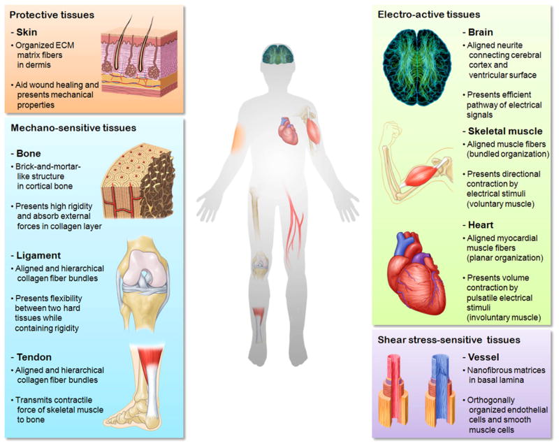
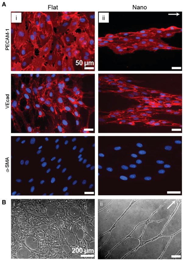
Human tissues are intricate ensembles of multiple cell types embedded in complex and well-defined structures of the extracellular matrix (ECM). The organization of ECM is frequently hierarchical from nano to macro, with many proteins forming large scale structures with feature sizes up to several hundred microns. Inspired from these natural designs of ECM, nanotopography-guided approaches have been increasingly investigated for the last several decades. Results demonstrate that the nanotopography itself can activate tissue-specific function in vitro as well as promote tissue regeneration in vivo upon transplantation. In this review, we provide an extensive analysis of recent efforts to mimic functional nanostructures in vitro for improved tissue engineering and regeneration of injured and damaged tissues. We first characterize the role of various nanostructures in human tissues with respect to each tissue-specific function. Then, we describe various fabrication methods in terms of patterning principles and material characteristics. Finally, we summarize the applications of nanotopography to various tissues, which are classified into four types depending on their functions: protective, mechano-sensitive, electro-active, and shear stress-sensitive tissues. Some limitations and future challenges are briefly discussed at the end.


Figures

Fig. 1
Fig. 1
Various nanostructures in the human body. The tissues are classified into four categories, namely protective, mechano-sensitive, electro-active and shear stress-sensitive tissues with respect to the tissue specific environment and functions.
Fig. 2
Fig. 2
(A) Scanning electron microscope images of collagen type VI network observed in human neonatal foreskin. (B) Electrospun type I collagen as a biomimetic nanofibrous extracellular matrix. (C–F) In vivo wound healing test at 1 and 4 weeks. In the 1-week collagen nanofiber group, surface debris disappeared and promoted proliferation of young capillaries and fibroblast was observed (E), while in the control group surface debris exists and dense infiltration of leukocytes and fibroblasts was observed, demonstrating promoted early stage skin wound healing with the assistance of nanofiber matrix (C). However, in the 4-week group, the healing processes were similar (D, F). A is reprinted with permission from ref. [14]. B–F are reprinted with permission from ref. [108].
Fig. 3
Fig. 3
(A) The nanostructures of cortical bone composed of plate-like mineral crystals with a dimension of 2–4 nm-thick and up to 100 nm in length. (B) Cross-sectional view of bone structure. The plate-like crystals are embedded in collagen matrix with the volume ratio of mineral to matrix on the order of 1:2, demonstrating stratified stacks with slightly dislocated centers. (C) Nanopit arrays fabricated by nanoimprint lithography with poly(methyl methacrylate) (PMMA). The feature size is 120 nm in diameter, 100 nm in depth, 300 nm center-to-center spacing with arrangement of hexagonal, square, displaced square (±50 nm from true center) and random. (D–E) Osteopontin (OPN) and osteocalcin (OCN) of osteoprogenitors after 21 days of culture. Dense aggregates and enhanced OPN and OCN levels were observed in displaced square 50 (±50 nm from true center ) substrate (fourth column of C, D and E). A is reprinted with permission from ref. [20]. B is reprinted with permission from ref. [19]. C, D and E are reprinted with permission from ref. [127].
Fig. 4
Fig. 4
(A) Scanning electron microscopy image of ex vivo myocardium of adult rat heart. Unidirectionally organized matrix fibers were observed. (B) Heart-inspired nanopatterned substratum made of polyethylene glycol (PEG) hydrogels. (C) Magnified view of well-aligned rat myocardium. (D) Scanning electron microscopy image of NRVMs cultured on anisotropically nanofabricated substratum, showing similar morphology to (C). (E) Contraction map of NRVM monolayers cultured on flat and nanopatterns. Unlike the random contraction on flat substratum, NRVMs cultured on anisotropic nanopattern demonstrated unidirectional contraction, which is similar to real heart. (F) Action potential propagation analysis of NRVMs cultured on flat and anisotropic nanopatterns. Point stimulation of 3 Hz in 0 ms (indicated by white arrows) propagated anisotropically on cells cultured on nanopatterns, while isotropic propagation was observed in the flat case. Reprinted with permission from ref. [167].
Fig. 5
Fig. 5
Supercellular band formation on flat and nanopatterned substrates after 6-day culture of endothelial cells (ECs). (A) Immunostaining images of typical endothelial markers including PECAM-1, VEcad and α-SMA. The direction of the arrow indicates topographical orientation. (B) In vitro capillary formation assay. Endothelial progenitor cells (EPCs) cultured on nanopatterns showed long and extensive network of capillary tube (ii), while on flat substrates unorganized and low density of capillary tubes were observed (i). Scale bar: 50 μm (A), 200 μm (B). Reprinted with permission from ref. [289].

References

    1. Messenger MP, Tomlins PE. Regenerative medicine: a snapshot of the current regulatory environment and standards. Adv Mater. 2011;23:H10–H17. - PubMed
    1. Mason C. Regenerative medicine 2.0. Regen Med. 2007;2:11–18. - PubMed
    1. Lo CM, Wang HB, Dembo M, Wang YL. Cell movement is guided by the rigidity of the substrate. Biophys J. 2000;79:144–152. - PMC - PubMed
    1. Carter SB. Principles of cell motility: the direction of cell movement and cancer invasion. Nature. 1965;208:1183–1187. - PubMed
    1. Zhao M, Song B, Pu J, Wada T, Reid B, Tai GP, Wang F, Guo AH, Walczysko P, Gu Y, Sasaki T, Suzuki A, Forrester JV, Bourne HR, Devreotes PN, McCaig CD, Penninger JM. Electrical signals control wound healing through phosphatidylinositol-3-OH kinase-gamma and PTEN. Nature. 2006;442:457–460. - PubMed

Publication types