Review
. 2012 Sep;18(9):534-43.
doi: 10.1016/j.molmed.2012.08.001. Epub 2012 Aug 23.

Molecular mechanisms mediating metastasis of hypoxic breast cancer cells

Affiliations
Review

Molecular mechanisms mediating metastasis of hypoxic breast cancer cells

Gregg L Semenza. Trends Mol Med. 2012 Sep.

Abstract

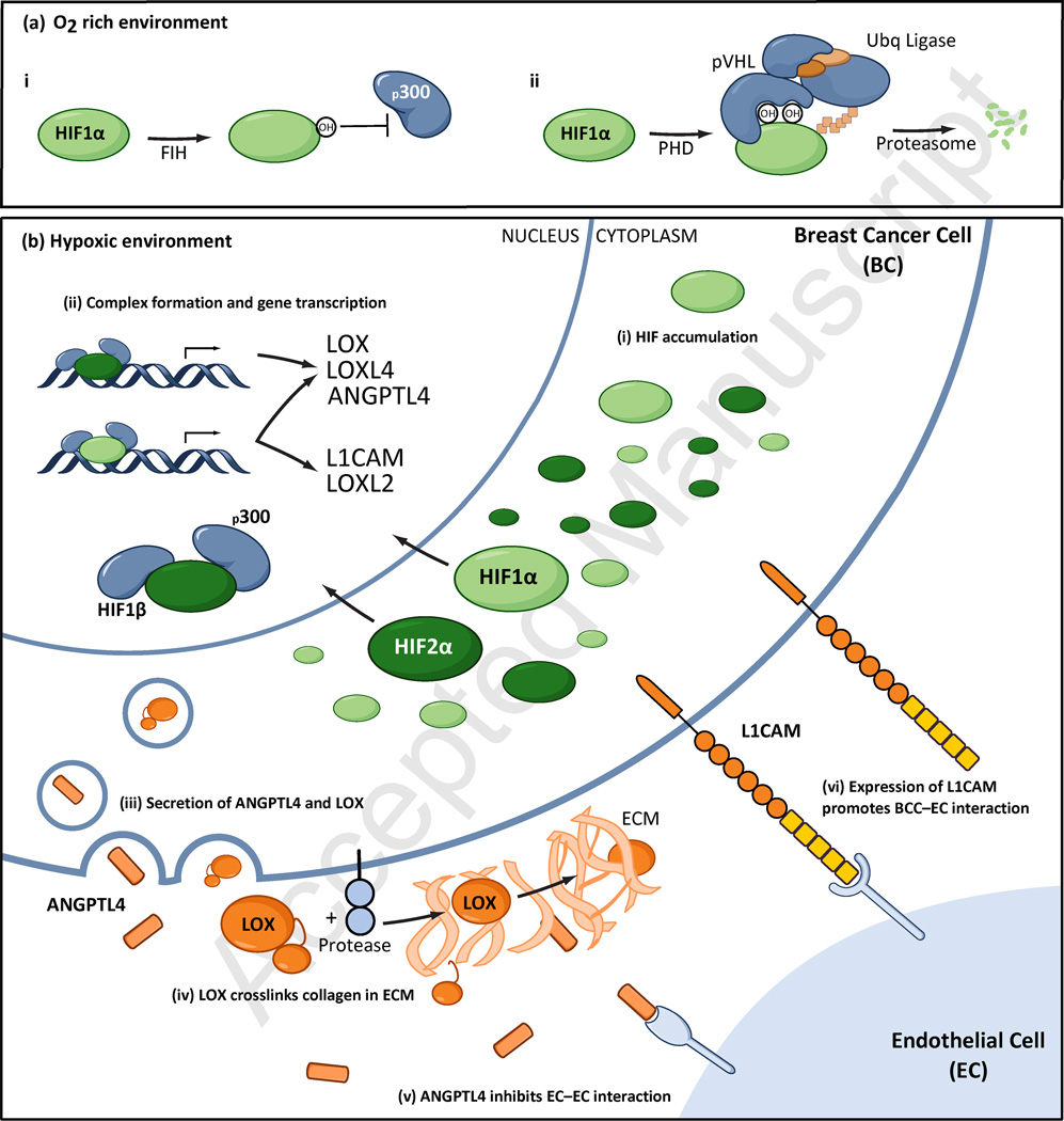
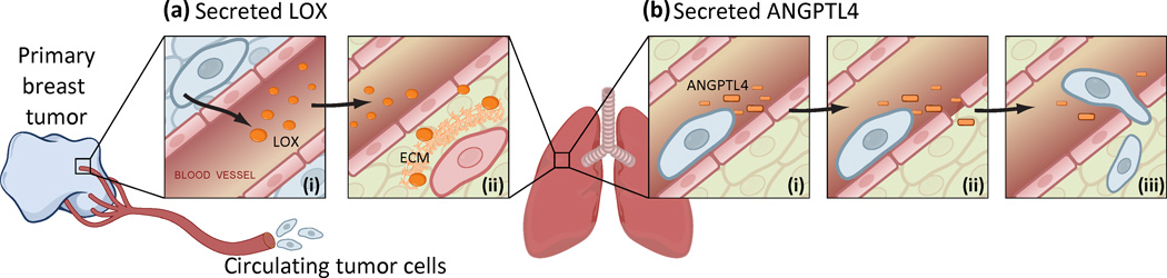
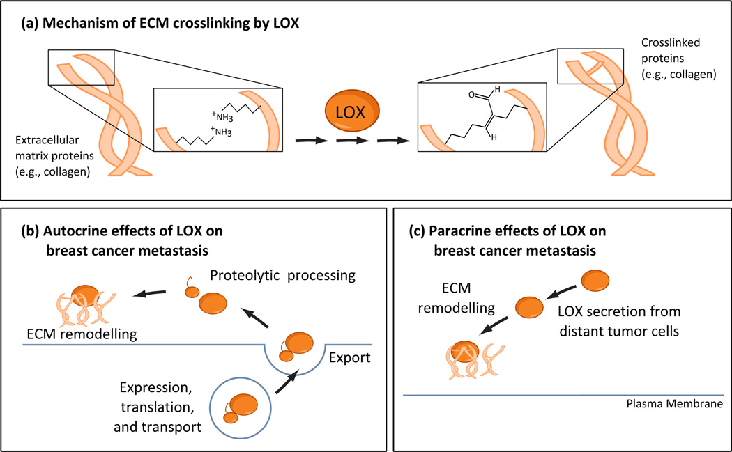
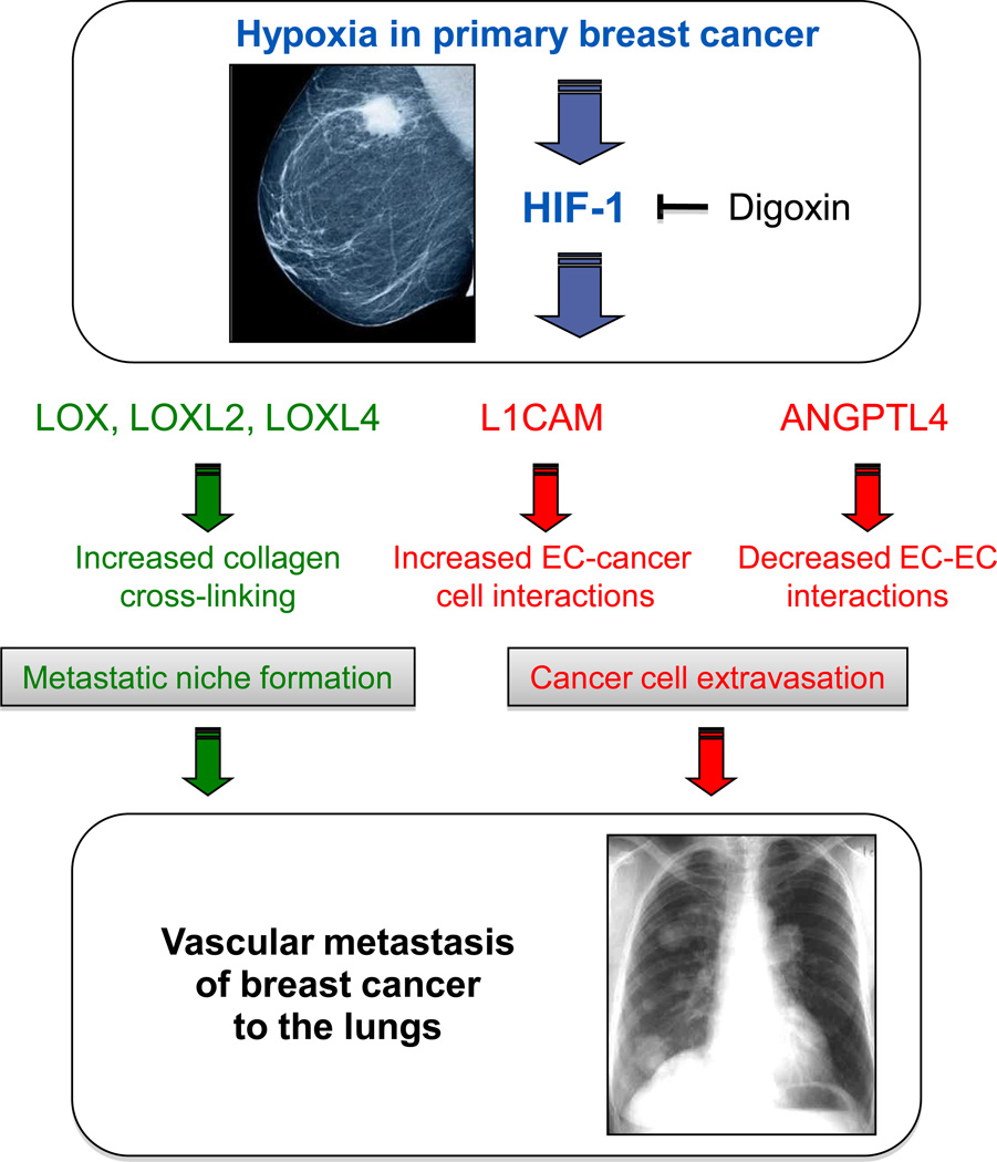
Breast cancers contain regions of intratumoral hypoxia in which reduced O(2) availability activates the hypoxia-inducible factors HIF-1 and HIF-2, which increase the transcription of genes encoding proteins that are required for many important steps in cancer progression. Recently, HIFs have been shown to play critical roles in the metastasis of breast cancer to the lungs through the transcriptional activation of genes encoding angiopoietin-like 4 and L1 cell adhesion molecule, which promote the extravasation of circulating cancer cells from the lung vasculature, and the lysyl oxidase family members LOX, LOXL2, and LOXL4, which promote invasion and metastatic niche formation. Digoxin, a drug that inhibits HIF-1 activity, blocks primary tumor growth, vascularization, invasion, and metastasis in ex vivo and in vivo assays.


Figures

Figure 1
Figure 1
Mechanisms and consequences of HIF activation in hypoxic breast cancer cells. (a) In well oxygenated cells, HIF-1α (and HIF-2α) are subjected to two O2-dependent hydroxylation reactions. (i) FIH-1 hydroxylates asparagine-803, which prevents HIF-1α from interacting with the coactivator p300 and renders it transcriptionally inactive. (ii) PHD2 hydroxylates proline-402 and/or proline-564, which allows HIF-1a to interact with VHL and renders it subject to ubiquination and proteasomal degradation. (b) In hypoxic cells, the hydroxylation reactions are inhibited, HIF-1a and HIF-2a accumulate, dimerize with HIF-1b, recruit p300, bind to target genes, and activate their transcription, leading to the expression of L1 cell adhesion molecule (L1CAM), angiopoietin-like 4 (ANGPTL4), lysyl oxidase (LOX), and LOX-like proteins 2 and 4 (LOXL2, LOXL4), which promote breast cancer metastasis by effects on endothelial cells (ECs) and the extracellular matrix (ECM).
Figure 2
Figure 2
Mechanisms by which L1CAM mediates cell–cell interactions. L1CAM-expressing cells have been reported to interact with other cells via homophilic interactions (a) or by heterophilic interactions with neuropilin 1 (NRP1), integrins (b), or CD24 (c).
Figure 3
Figure 3
Pro-metastatic effects of proteins secreted by hypoxic breast cancer cells. Breast cancer cells in the primary tumor are subjected to hypoxia, which induces the expression of LOX and ANGPTL4. (a) In the primary tumor, LOX is secreted into the circulation (i) and crosslinks collagen in the lungs, leading to the recruitment of bone marrow-derived cells that together define the pre-metastatic nice (ii). (b) A circulating tumor cell from the hypoxic primary tumor that has adhered to endothelial cells (ECs) in a pulmonary blood vessel secretes ANGPTL4, which inhibits EC–EC interaction, thereby facilitating the extravasation of the breast cancer cells into the lung parenchyma.
Figure 4
Figure 4
Local and distant effects of LOX. (a) LOX (and LOXL proteins) crosslink collagen proteins, thereby altering the physical properties of the ECM. (a) Crosslinking of ECM proteins by LOX in the primary tumor promotes local cancer cell invasion. (b) Secretion of LOX into the circulation results in ECM remodeling at distant sites such as the lungs, which promotes metastasis.
Figure 5
Figure 5
Molecular and cellular mechanisms by which HIF-1 promotes metastasis of hypoxic breast cancer to the lungs. Hypoxia in the primary tumor induces the activity of HIF-1 (and HIF-2) in breast cancer cells, leading to increased transcription of genes encoding proteins that subsequently promote metastasis. The expression of L1CAM on the cell surface of circulating breast cancer cells increases their adherence to the endothelial cells (ECs) of pulmonary blood vessels (margination), whereas the secretion of ANGPTL4 decreases EC–EC interactions, which promotes the extravasation of breast cancer cells into the lung parenchyma. Prior to the arrival of breast cancer cells, a pre-metastatic niche forms in the lung through the activity of LOX/LOXL proteins, which are secreted by hypoxic breast cancer cells in the primary tumor and crosslink collagen fibers in the lung, thereby promoting the retention of bone marrow-derived cells, which in turn promote the recruitment of metastatic breast cancer cells. Digoxin inhibits the synthesis of HIF-1α (and to a lesser extent, HIF-2α) and thereby blocks all of the downstream steps described above.
Figure I
Figure I
Functions of L1CAM observed in other cell types
Figure II
Figure II
Functions of LOX observed in other cell types

Similar articles

Cited by

References

    1. Vaupel P, et al. Tumor hypoxia and malignant progression. Methods Enzymol. 2004;381:335–354. - PubMed
    1. Vaupel P, et al. Detection and characterization of tumor hypoxia using pO2 histography. Antioxid. Redox Signal. 2007;9:1221–1235. - PubMed
    1. Vaupel P, et al. Prognostic potential of the pre-therapeutic tumor oxygenation status. Adv. Exp. Med. Biol. 2009;645:241–246. - PubMed
    1. Wilson WR, Hay MP. Targeting hypoxia in cancer therapy. Nat. Rev. Cancer. 2011;11:393–410. - PubMed
    1. Semenza GL. Hypoxia inducible factors in physiology and medicine. Cell. 2012;148:399–408. - PMC - PubMed

Publication types

MeSH terms

Substances