. 2012 Nov 5;363(1-2):111-21.
doi: 10.1016/j.mce.2012.08.005. Epub 2012 Aug 16.

Progestin modulates the lipid profile and sensitivity of breast cancer cells to docetaxel

Affiliations

Progestin modulates the lipid profile and sensitivity of breast cancer cells to docetaxel

Isabel R Schlaepfer et al. Mol Cell Endocrinol. .

Abstract

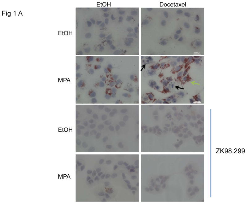
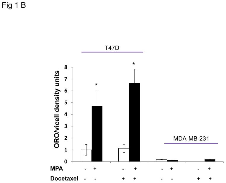
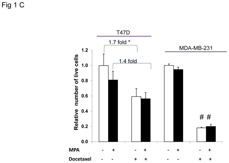
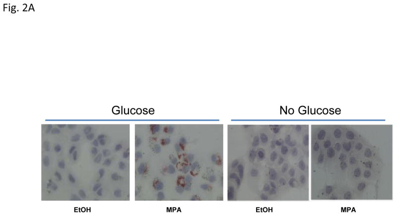
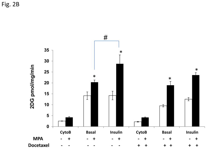
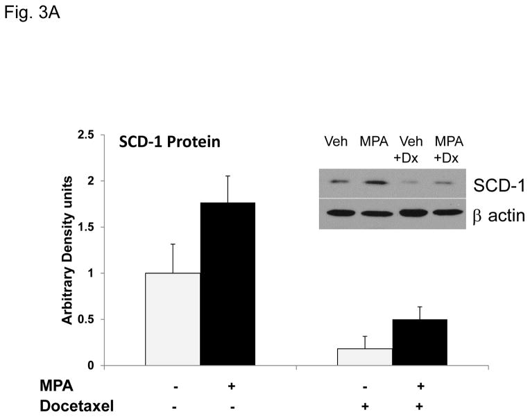
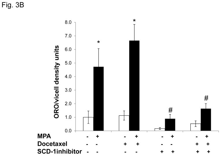
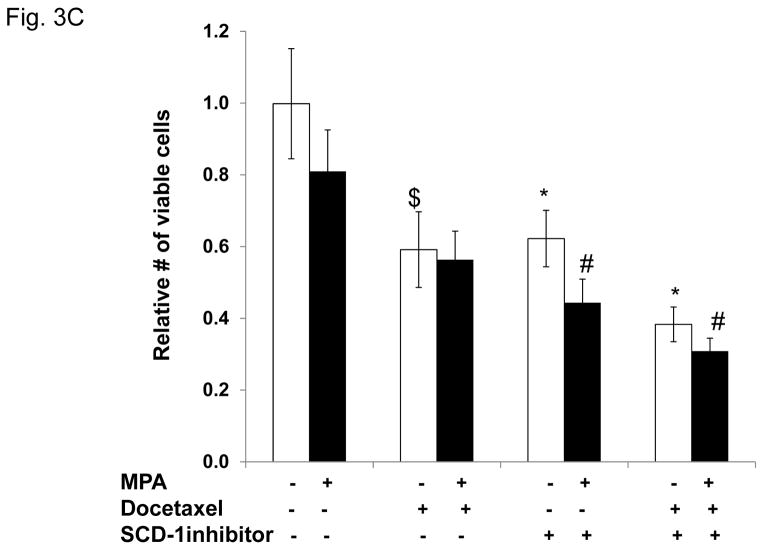
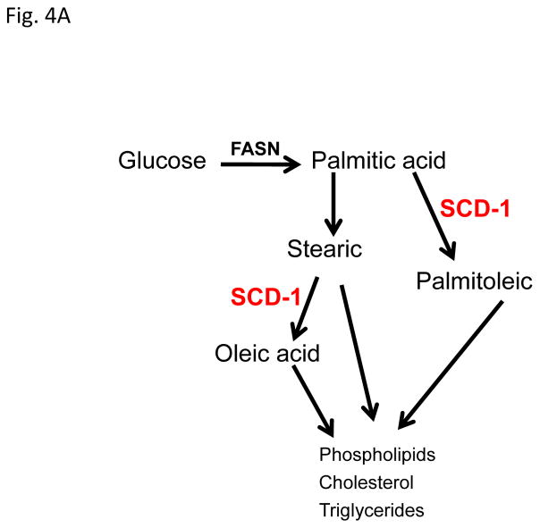
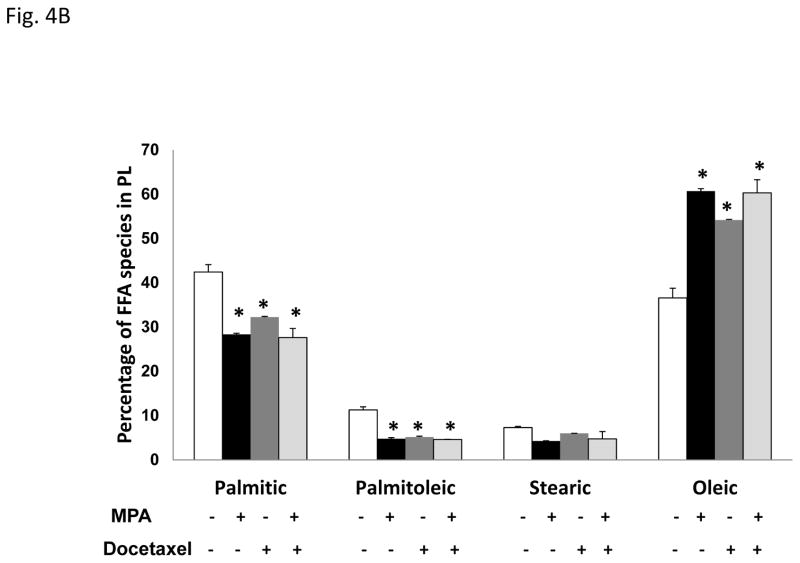
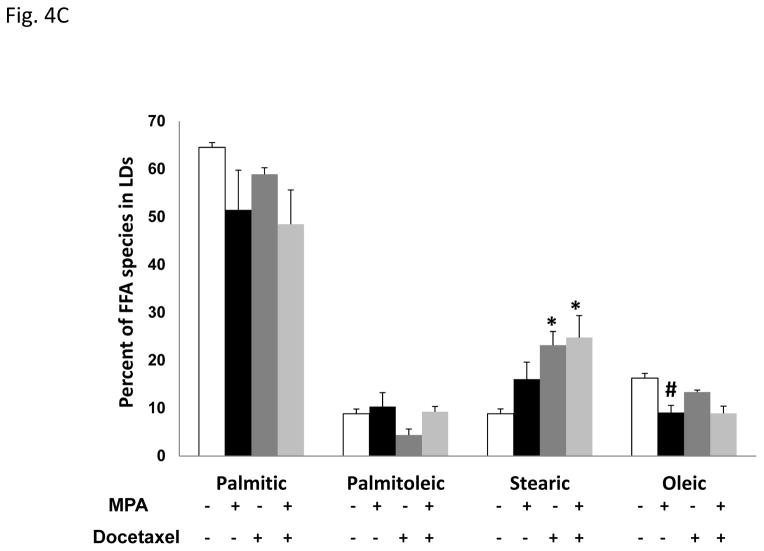
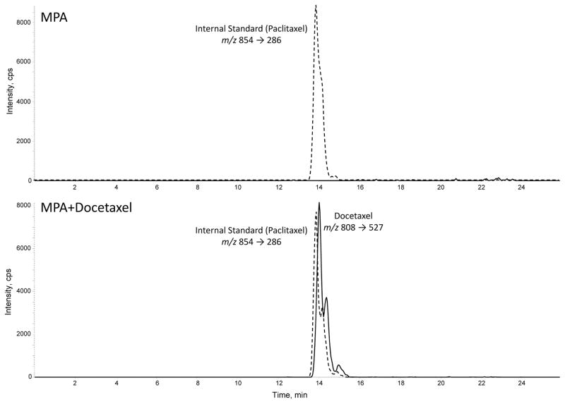
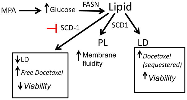
Progestins induce lipid accumulation in progesterone receptor (PR)-positive breast cancer cells. We speculated that progestin-induced alterations in lipid biology confer resistance to chemotherapy. To examine the biology of lipid loaded breast cancer cells, we used a model of progestin-induced lipid synthesis. T47D (PR-positive) and MDA-MB-231 (PR-negative) cell lines were used to study progestin response. Oil red O staining of T47D cells treated with progestin showed lipid droplet formation was PR dependent, glucose dependent and reduced sensitivity to docetaxel. This protection was not observed in PR-negative MDA-MB-231 cells. Progestin treatment induced stearoyl CoA desaturase-1 (SCD-1) enzyme expression and chemical inhibition of SCD-1 diminished lipid droplets and cell viability, suggesting the importance of lipid stores in cancer cell survival. Gas chromatography/mass spectroscopy analysis of phospholipids from progestin-treated T47D cells revealed an increase in unsaturated fatty acids, with oleic acid as most abundant. Cells surviving docetaxel treatment also contained more oleic acid in phospholipids, suggesting altered membrane fluidity as a potential mechanism of chemoresistance mediated in part by SCD-1. Lastly, intact docetaxel molecules were present within progestin induced lipid droplets, suggesting a protective quenching effect of intracellular lipid droplets. Our studies suggest the metabolic adaptations produced by progestin provide novel metabolic targets for future combinatorial therapies for progestin-responsive breast cancers.


Figures

Figure 1
Figure 1. Progestin induces lipid accumulation and increases chemoresistance in PR+ breast cancer cells
A, representative photographs of Oil red O stained T47D cells grown on coverslips and treated with or without MPA for 6 days. Docetaxel or ethanol (EtOH) was added for the last 2 days of treatment. Green arrow shows viable cell following docetaxel treatment, black arrows denote apoptotic cells. Lower panels: samples treated as above in the presence of the antiprogestin, ZK98,299. Lipid staining is absent in the presence of the anti-progestin. B, Quantification of Oil red O staining photographs of T47D and MDA-MB-231 cells using arbitrary density units. White and black bars represent ethanol and MPA treatments respectively. * p< 0.05, MPA versus vehicle for each cell line. Bars represent mean ±SD. Experiments were performed 5 times in independent triplicate assays. C, Number of viable T47D and MDA-MB-231 cells after MPA and docetaxel treatments as indicated above. Relative cell viability is shown for each cell line normalized to the untreated control.* p=0.02 vehicle vs. docetaxel for each treatment group (vehicle or MPA). # p< 0.001 vehicle versus docetaxel for each treatment group. Experiments were performed 7 times in independent triplicate assays.
Figure 2
Figure 2. Glucose uptake from the media is increased in progestin-treated cells and required for lipid droplet formation
A, Photographs of Oil red O stains of T47D cells treated with vehicle or MPA with or without glucose. B, Glucose uptake of T47D cells treated with ethanol (white bars) or MPA (black bars) for 6 days. The last 2 days of treatment, cells were exposed to docetaxel where indicated. Cytochalasin-B (Cyto-B) was used to block glucose transporters and reveal the transporter-mediated glucose uptake. Bars represent mean ±SD. Statistically significant values between ethanol and MPA treatment are indicated: * p < 0.01, difference between ethanol (open bars) and MPA. # p=0.006, difference between MPA and MPA+insulin treatments. Experiments were performed three independent times in duplicate and one representative experiment is shown.
Figure 3
Figure 3. Progestin-mediated SCD-1 expression is required for lipid droplet formation and viability of T47D exposed to docetaxel
A, Densitometry of two independent immunoblots for SCD-1 protein normalized to beta-actin is shown. Cells were treated as indicated. Inset; immunoblot for SCD-1 with β-actin as a loading control. B, Quantification of Oil red O staining of T47D cells after MPA and docetaxel treatments with or without SCD-1 inhibitor. White and black bars represent ethanol and MPA treatments respectively. * p< 0.05, MPA versus vehicle. # p ≤ 0.006 compared to MPA+docetaxel. Bars represent mean ±SEM of 5 independent assays in triplicate. C, Number of viable T47D cells after MPA and docetaxel treatments as indicated above. Relative cell viability is shown normalized to the untreated control. $ p=0.02 compared to vehicle alone. * p ≤ 0.005 compared to vehicle alone. # p ≤ 0.006 compared to MPA alone. Experiments were performed 7 times in independent triplicate assays.
Figure 4
Figure 4. Progestin alters the lipid profile of T47D cells
A, Diagram of the site of action of the SCD-1 enzyme. B, Gas Chromatography-Mass Spectroscopy (GCMS) analysis of most common fatty acids in the T47D phospholipid fraction: Palmitic (16C:0), palmitoleic (16C:1), stearic (18C:0) and oleic acid (18C:1). Bars represent mean ±SD. * p < 0.01, docetaxel, MPA or MPA+docetaxel treatment versus vehicle. C, GCMS of triglyceride fractions (lipid droplets) of T47D lipid extracts. Bars represent mean ±SD. * p ≤ 0.04, docetaxel or MPA+docetaxel treatment versus vehicle. # p = 0.01, vehicle versus MPA or MPA+docetaxel treatment.
Figure 5
Figure 5. Liquid chromatography-tandem mass spectroscopy (LCMS/MS) analysis of docetaxel from isolated lipid droplets of T47D cells
Paclitaxel was used as a reference molecule for the analysis. Total lipids from isolated lipid droplets of cells treated with MPA+vehicle (ethanol) or MPA+docetaxel (100nM) were extracted using solid-phase cartridges and analyzed by LC/MS/MS. The chromatograms show the ion intensity profiles for docetaxel (solid line) and paclitaxel (dashed line).
Figure 6
Figure 6. Diagram of the proposed mechanism of progestin action in the lipid profile and survival of T47D cells exposed to docetaxel
SCD-1 increases membrane fluidity and induces the formation of lipid droplets that are able to sequester active docetaxel molecules, favoring cell survival. SCD-1 inhibition leads to lack of lipid droplets, increased free docetaxel and decreased cell viability as well as increases membrane fluidity in the FASN, fatty acid synthase. SCD-1, steroyl CoA desaturase. PL, phospholipid. LD, lipid droplets.

Similar articles

Cited by

References

    1. Badtke MM, Jambal P, Dye WW, Spillman MA, Post MD, Horwitz KB, Jacobsen BM. Unliganded progesterone receptors attenuate taxane-induced breast cancer cell death by modulating the spindle assembly checkpoint. Breast Cancer Res Treat. 2012;131:75–87. - PMC - PubMed
    1. Baritaki S, Apostolakis S, Kanellou P, Dimanche-Boitrel MT, Spandidos DA, Bonavida B. Reversal of tumor resistance to apoptotic stimuli by alteration of membrane fluidity: therapeutic implications. Adv Cancer Res. 2007;98:149–190. - PubMed
    1. Bayet-Robert M, Morvan D, Chollet P, Barthomeuf C. Pharmacometabolomics of docetaxel-treated human MCF7 breast cancer cells provides evidence of varying cellular responses at high and low doses. Breast Cancer Res Treat 2009 - PubMed
    1. BLIGH EG, DYER WJ. A rapid method of total lipid extraction and purification. Can J Biochem Physiol. 1959;37:911–917. - PubMed
    1. Chajes V, Hulten K, Van Kappel AL, Winkvist A, Kaaks R, Hallmans G, Lenner P, Riboli E. Fatty-acid composition in serum phospholipids and risk of breast cancer: an incident case-control study in Sweden. Int J Cancer. 1999;83:585–590. - PubMed

Publication types

MeSH terms