. 2012 Dec 6:225:269-82.
doi: 10.1016/j.neuroscience.2012.08.033. Epub 2012 Aug 23.

Ectopic uterine tissue as a chronic pain generator

Affiliations

Ectopic uterine tissue as a chronic pain generator

P Alvarez et al. Neuroscience. .

Abstract

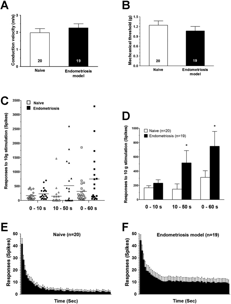
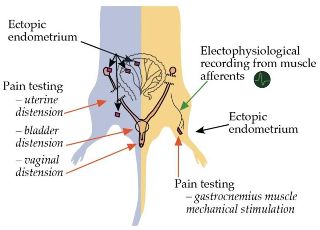
While chronic pain is a main symptom in endometriosis, the underlying mechanisms and effective therapy remain elusive. We developed an animal model enabling the exploration of ectopic endometrium as a source of endometriosis pain. Rats were surgically implanted with autologous uterus in the gastrocnemius muscle. Within two weeks, visual inspection revealed the presence of a reddish-brown fluid-filled cystic structure at the implant site. Histology demonstrated cystic glandular structures with stromal invasion of the muscle. Immunohistochemical studies of these lesions revealed the presence of markers for nociceptor nerve fibers and neuronal sprouting. Fourteen days after surgery rats exhibited persistent mechanical hyperalgesia at the site of the ectopic endometrial lesion. Intralesional, but not contralateral, injection of progesterone was dose-dependently antihyperalgesic. Systemic administration of leuprolide also produced antihyperalgesia. In vivo electrophysiological recordings from sensory neurons innervating the lesion revealed a significant increase in their response to sustained mechanical stimulation. These results are consistent with clinical and pathological findings observed in patients with endometriosis, compatible with the ectopic endometrium as a source of pain. This model of endometriosis allows mechanistic exploration at the lesion site facilitating our understanding of endometriosis pain.


Conflict of interest statement

Competing financial interests

The authors declare that no conflict of interest exists in connection with this study.

Figures

Figure 1
Figure 1
Morphologic and behavioral changes induced by ectopic uterine tissue. (A) 3 weeks after surgical implant of uterus tissue on the gastrocnemius muscle, surgical dissection reveals the gross anatomy of the cystic lesion and close muscular structures. (B) Microscopic analysis of cystic lesion reveals skeletal muscle invasion by ectopic endometrium and development of accessory cystic structures. Hematoxylin-eosin staining, 50X. (C) High magnification detail of panel B showing embedding of uterine tissue in skeletal muscle; Hematoxylin-eosin staining, 100X. (D) Mechanical hyperalgesia was already present at day 10 post-implantation after unilateral transplantation of endometrium, remaining undiminished for the duration of the testing period 27 days post-implantation. Hyperalgesia was significantly greater than in rats implanted with adipose tissue on to gastrocnemius muscle. (E) Mechanical hyperalgesia at the site of cystic lesion was evaluated at proestrus, estrus, metestrus and diestrus. No significant difference in nociceptive threshold was observed after testing ipsilateral or contralateral to the implant. (F) Intralesional administration of progesterone (P4) at doses of 1 µg, or (G) 3 µg at the site of the endometrial implant significantly attenuated mechanical hyperalgesia (repeated measures one-way ANOVA with Dunnett’s multiple comparison test). (H, I) The same doses of progesterone had no effect on nociceptive mechanical threshold when injected in the contralateral side to the uterus implant. Behavioral data are presented as % of change of baseline values. **P < 0.01; ***P < 0.001.
Figure 2
Figure 2
Systemic administration of leuprolide modulates the mechanical hyperalgesia induced by ectopic endometrium. The s.c. administration of leuprolide produced a bi-phasic effect on mechanical hyperalgesia associated to endometriosis-like lesions: one day after injection such hyperalgesia was increased; then a progressive attenuation of the hyperalgesia was observed, which persisted at least until day 7 after leuprolide injection. *P < 0.05; **P < 0.01; ***P < 0.001.
Figure 3
Figure 3
Comparative histologic study of eutopic endometrium and ectopic endometrial implants. (A) Rat eutopic uterus sample showing vimentin reactivity in endometrial stromal fibroblasts and unlabeled endometrial surface epithelium and glands (arrows). (B) Rat eutopic uterus sample showing cytokeratin 18 reactivity in endometrial surface epithelium and glands and lack of immunolabeling in endometrial stromal fibroblasts. (C) Uterine implant showing vimentin reactivity in endometrial stromal fibroblasts and endomysium, and non immuno-labeled skeletal muscle fibers. (D) Cystic uterine implant showing cytokeratin 18 reactivity in the endometrial epithelium and non immuno-labeled skeletal muscle fibers. (E) Hematoxylin-eosin staining showing cystic structures (indicated by *) and endometrial stroma embedded into the skeletal muscle tissue. (F) Serial section from the same specimen in panel e showing vimentin positive elements infiltrating between the skeletal muscle fibers. (G) Low magnification of implant showing extensive invasion of muscle by vimentin-positive endometrial stromal fibroblasts. (H) High magnification detail of panel G showing vimentin-positive endometrial fibroblasts deeply invading endomysium and causing displacement and compression of the skeletal muscle fibers. Original magnifications 50X (G) or 100X (A-F, H).
Figure 4
Figure 4
Innervation of cystic lesions by muscle nociceptor and its axonal sprouts. Gastrocnemius muscle and ectopic uterine implant cyst immuno-labeled with antibodies against CGRP (A) and counterstained with DAPI (B, C); white arrows indicate nerve fiber-like structures. Gastrocnemius muscle and ectopic uterine implant cyst labeled with the IB4 (D) and counterstained with DAPI (E, F); white arrows indicate nerve fiber-like structures. (G) Immunolabeling for the marker for neuronal sprouting GAP43 is observed in the cyst wall. (H) Dot-like structures in the cyst wall and a nerve fiber-like structure surrounding the cyst are immune-positive to CGRP. (I) Merged image shows that only dot-like structures in the cyst wall are co-labeled for GAP43 and CGRP. The cyst lumen is indicated by *. Scale bars represent 100 µm.
Figure 5
Figure 5
Sensitized nociceptors innervating ectopic uterine tissue. Two weeks after surgery, C-fiber uterine tissue implants, did not have a significant change in (A) mechanical threshold, or (B) conduction velocity. In contrast, they had significantly enhanced (C, D) C-fiber response to sustained mechanical stimulation (60 s), as revealed by increased number of spikes in recordings obtained during early (10 s) and late (50 s) parts of the stimulation period compared to control values obtained from naïve female rats. This was also evident in the time-course histograms of the C-fiber response, representing recordings obtained in naïve-control (E) and implanted (F) rats. Comparisons between naïve and implanted female rats were made using one-tail Student’s t-test for paired samples with Welch’s correction; *P < 0.05.
Figure 6
Figure 6
A-type and DR-type potassium currents are not altered in cyst-innervating DRG neurons. Examples of retrogradelly-labeled DRG cells with DiI (shown in red) and IB4-FITC labeling (shown in green) in culture: (A) a DiI labeled, IB4-negative DRG cell; (B) a double labeled (DiI labeled-IB4-positive) DRG cell. (C) Current-voltage curves (normalized to cell capacitance) representing A-type (sensitive to 3,4-diaminopyridine, DAP) current densities are shown. The peak current density in endometriosis DRG neurons (open circles, n=7) was not significantly different to that of control neurons (closed squares, n=7). (D) Representative current traces obtained from DRG neurons of control rats and rats submitted to the endometriosis pain model in the presence of DAP. (E) The DR-type potassium current-density curves obtained in the presence of tetraethylammonium (TEA) were not significantly different in endometriosis DRG neurons (open circles, n=8) compared to control (closed squares, n=7). (F) Representative current traces obtained from DRG neurons of control rats and rats submitted to the endometriosis pain model in the presence of TEA.
Figure 7
Figure 7
Comparison of rat models of endometriosis pain. The rat model developed by Vernon and Wilson (1985) and adapted by Berkley and colleagues (2001) for the behavioral assessment of endometriosis pain (left side of the figure), differs in several ways from the model presented here (right side of the figure). In the former model of endometriosis pain, ectopic uterine tissue is implanted in the peritoneal cavity and nociceptive testing performed by mechanical stimulation of non-implanted structures (i.e., uterine, bladder or vaginal distension). In contrast, the model presented here allows direct nociceptive testing of uterine tissue-implanted gastrocnemius muscle, as well as, electrophysiological recording from nociceptive afferents innervating the ectopic uterine tissue and local testing of putative modulatory agents. Additional advantages include: (1) easy access for retrograde labeling of DRG neurons innervating from endometriosis-like lesions allowing for morphological and in vitro electrophysiological studies and, (2) contralateral assessment of nociceptive responses or drug administration in order to evaluate for extra-local effects of putative modulators.

References

    1. Alvarez P, Levine JD, Green PG. Eccentric exercise induces chronic alterations in musculoskeletal nociception in the rat. Eur J Neurosci. 2010;32:819–825. - PMC - PubMed
    1. Benowitz LI, Routtenberg A. GAP-43: an intrinsic determinant of neuronal development and plasticity. Trends Neurosci. 1997;20:84–91. - PubMed
    1. Berkley KJ, Cason A, Jacobs H, Bradshaw H, Wood E. Vaginal hyperalgesia in a rat model of endometriosis. Neurosci Lett. 2001;306:185–188. - PubMed
    1. Berkley KJ, Rapkin AJ, Papka RE. The pains of endometriosis. Science. 2005;308:1587–1589. - PubMed
    1. Botha AJ, Halliday AE, Flanagan JP. Endometriosis in gluteus muscle with surgical implantation. A case report. Acta Orthop Scand. 1991;62:497–499. - PubMed

Publication types

MeSH terms