. 2012 Nov 1;371(1):66-76.
doi: 10.1016/j.ydbio.2012.08.006. Epub 2012 Aug 18.

Two functional domains in C. elegans glypican LON-2 can independently inhibit BMP-like signaling

Affiliations

Two functional domains in C. elegans glypican LON-2 can independently inhibit BMP-like signaling

Suparna Taneja-Bageshwar et al. Dev Biol. .

Abstract

Glypicans are multifunctional proteoglycans with regulatory roles in several intercellular signaling pathways. Here, we examine the functional requirements for glypican regulation of bone morphogenetic protein (BMP)-mediated body length in C. elegans. We provide evidence that two parts of C. elegans glypican LON-2 can independently inhibit BMP signaling in vivo: the N-terminal furin protease product and the C-terminal region containing heparan sulfate attachment sequences. While the C-terminal protease product is dispensable for LON-2 minimal core protein activity, it does affect the localization of LON-2. Cleavage of LON-2 into two parts at the conserved furin protease site is not required for LON-2 to inhibit BMP-like signaling. The glycosyl-phosphatidylinositol (GPI) membrane anchor is also not absolutely required for LON-2 activity. Finally, we show that an RGD protein-protein interaction motif in the LON-2 N-terminal domain is necessary for LON-2 core protein activity, suggesting that LON-2 inhibits BMP signaling by acting as a scaffold for BMP and an RGD-binding protein.


Figures

Fig. 1
Fig. 1
The LON-2 core protein is active in vivo. Schematic illustrations and in vivo activity of LON-2-derived constructs are presented. The positions of LON-2 elements are shown: N-terminal signal sequence at amino acids 1–18, gray block; 14 conserved cysteines, solid blue lines; RGD at amino acids 348–350, light blue line; RLGR consensus furin protease recognition site at amino acids 365–368, dashed maroon line; HS GAG attachment sequences at amino acids 374–375 and 442–445, dotted red lines; and a GPI linkage site at the C-terminus at amino acid 489, black block. The schematic representation of LON-2 is used for Figs. 1, 3–5. All constructs are assayed for LON-2 activity by quantitating their ability to reduce the long body length defect of lon-2(e678) animals. For Figs. 1, 3–5, the body length average of staged adult animals of each transgenic strain was compared to identically staged lon-2(e678) animals ± 95% confidence interval, where the lon-2(e678) body length average is normalized to 100%. Two strains and at least 30 transgenic animals per strain were measured for each LON-2 construct. The lon-2(e678) body size phenotype-rescuing activity of each LON-2 construct is noted in the last column. First construct, LON-2(GFP): full length, GFP-tagged LON-2. Second construct, LON-2(GAG): single LON-2(GFP) GAG-binding site variant S374A. Third construct, LON-2(1,2GAG): double LON-2(GFP) GAG-binding site variant S442A S444A. Fourth construct, LON-2(1,2,3GAG): triple LON-2(GFP) GAG-binding site variant S374A S442A S444A, which lacks all consensus HS attachment sites. Wild-type strain (N2) results are provided for comparison.
Fig. 2
Fig. 2
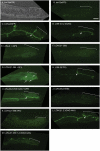
Cell membrane localization of GFP-tagged LON-2 constructs depends on the presence of a GPI anchor and the LON-2 C-terminus. Differential Interference Contrast (DIC) (A) or spinning disk confocal (B-M) images were taken of the posterior intestine of L4 stage hermaphrodites using a 63 × objective and 2.5 or 5.0 s exposure. Bar in (B)= 10 μm. As a common landmark, the most posterior dorsal intestinal cell (int9R) is bracketed in each image. Short white arrows mark the cell–cell junction of the most posterior dorsal intestinal cells (int8R and int9R) with the most posterior ventral intestinal cell (int9L) (C, D, G–I, L). (A) DIC view of posterior intestine seen in (B). 1.0 s exposure. (B) Non-transgenic lon-2(e678) animal shows some background gut autofluorescence. 5.0 s exposure. (C) LON-2(GFP), GFP-tagged full-length LON-2. 2.5 s exposure. (D) LON-2(1,2,3GAG), LON-2 lacking all GAG-attachment sites. 2.5 s exposure. (E) LON-2(ΔGPI), soluble LON-2 lacking sequence to attach the GPI anchor. 5.0 s exposure. (F) LON-2(1-368), the soluble N-terminal protease product of LON-2. 5.0 s exposure. (G) LON-2(1-364 +GPI), an N-terminal portion of LON-2 that is membrane-tethered by GPI, is weakly visible at cell junctions (short white arrow) but appears to localize primarily to the cytoplasm and clusters on the cell membrane (long white arrows). 5.0 s exposure. (H) LON-2(FCS), full-length LON-2 lacking a furin cleavage site. 2.5 s exposure. (I) LON-2(423-508 GAG +GFP), a C-terminal portion of LON-2 lacking GAG attachment sites. 2.5 s exposure. (J) LON-2(423-488), a C-terminal portion of glycanated LON-2 that lacks the GPI attachment sequence. 5.0 s exposure. (K) LON-2(1-368 AAA), an R348GD-to-AAA variant derived from soluble LON-2(1-368). 5.0 s exposure. (L) LON-2(1,2,3GAG AAA), full-length LON-2 lacking GAG-attachment sites and the RGD motif. 2.5 s exposure. (M) LON-2(1-488 1,2,3,GAG AAA), full-length LON-2 lacking GAG-attachment sites, the RGD motif, and GPI anchor sequence. 5.0 s exposure. Although the signals from the LON-2 variants appear to vary in intensity, these results varied between animals within strains and may not be physiologically relevant.
Fig. 3
Fig. 3
Soluble LON-2 is sufficient but not necessary for activity in vivo. First construct, LON-2(ΔGPI): LON-2(GFP) variant with the GPI attachment site (amino acids 489–508) deleted. Second construct, LON-2(1-368): LON-2(GFP) variant comprising the N-terminal protease product sequence. Third construct, LON-2(1-364 + GPI): LON-2(1-368) variant that lacks a furin protease site and contains the LON-2 GPI anchor sequence at the C-terminus. Fourth construct, LON-2(FCS): LON-2(GFP) variant R365LGR to A365LGA, which lacks a furin protease site. Body length-restoring activity is compared to lon-2(e678) animals ±95% confidence interval, where lon-2(e678) is 100%. Two strains and at least 30 animals per strain were measured for each LON-2 construct. LON-2(GFP) results are provided for comparison and represent independent measurements of one strain that were collected in the same trial sets as other results presented in this figure.
Fig. 4
Fig. 4
The glycanated, GPI-linked LON-2 C-terminus contains LON-2 activity and C. elegans glypican GPN-1 does not inhibit DBL-1 signaling in vivo. First construct, LON-2(423-508): LON-2 variant with the LON-2 secretion signal attached to the C-terminal 86-amino acid portion of LON-2. Second construct, LON-2(423-508 GAG): LON-2(423-508) variant that lacks both consensus HS attachment sites in this region. Third construct, LON-2(423-508 GAG + GFP): LON-2(423-508 GAG) variant with GFP sequence inserted between the secretion signal and the C-terminus sequences. Fourth construct, LON-2(423-488): LON-2(423-508) variant that lacks sequence to attach the GPI anchor. Fifth construct, GPN-1: full-length gpn-1 cDNA product. Annotations are the same used for labeling LON-2 variants. Body length-restoring activity is compared to lon-2(e678) animals ±95% confidence interval, where lon-2(e678) is 100%. Two strains and 30 animals per strain were measured for each LON-2 construct. LON-2(GFP) results are provided for comparison and represent independent measurements of one strain that were collected in the same trial sets as other results presented in this figure.
Fig. 5
Fig. 5
Soluble LON-2 protein core requires its RGD motif for in vivo activity. First construct, LON-2(1-347): LON-2(GFP) variant that contains amino acids 1-347, which lacks RGD sequence. Second construct, LON-2(1-368 AAA): LON-2(1-368) variant R348GD-to-AAA. Third construct, LON-2(1,2,3GAG AAA): LON-2(1,2,3 GAG) variant R348GD-to-AAA, which is a full length construct lacking GAG-attachment sites and the RGD motif. Fourth construct, LON-2(1-488 1,2,3GAG AAA): LON-2(1,2,3GAG AAA) variant that lacks sequence to attach the GPI anchor. Body length-restoring activity is compared to lon-2(e678) animals ±95% confidence interval, where lon-2(e678) is 100%. At least two strains and 30 animals per strain were measured for each LON-2 construct. LON-2(1-368) results presented in Fig. 3 are provided for comparison.
Fig. 6
Fig. 6
Model of LON-2 parts that regulate DBL-1/BMP signaling. Two parts of LON-2 can independently control DBL-1 signaling, the cylindrical N-terminal core protein within the first 368 amino acids (LON-1(1-368), middle), and the unstructured, GPI-anchored C terminal 86 amino acid region containing HS attachment sites (LON-2(423-508), right). Three arrowed elements are essential for the activity of either the N-terminal or C-terminal parts, but are not critical for the activity of full-length LON-2 (wild-type LON-2, left): the RGD protein-protein interaction motif (light blue) in the N-terminus, and the HS attachment sites (red) and GPI lipid anchor to the plasma membrane (thin black lines) in the C-terminus. This diagram is based on tertiary structure predictions from crystal structures of Dally-like and GPC-1 (Kim et al., 2011; Svensson et al., 2012).

Similar articles

Cited by

References

    1. Akiyama T, Kamimura K, Firkus C, Takeo S, Shimmi O, Nakato H. Dally regulates Dpp morphogen gradient formation by stabilizing Dpp on the cell surface. Dev. Biol. 2008;313:408–419. - PMC - PubMed
    1. Baeg GH, Lin X, Khare N, Baumgartner S, Perrimon N. Heparan sulfate proteoglycans are critical for the organization of the extracellular distribution of Wingless. Development. 2001;128:87–94. - PubMed
    1. Bernfield M, Gotte M, Park PW, Reizes O, Fitzgerald ML, Lincecum J, Zako M. Functions of cell surface heparan sulfate proteoglycans. Annu. Rev. Biochem. 1999;68:729–777. - PubMed
    1. Brandan E, Carey DJ, Larrain J, Melo F, Campos A. Synthesis and processing of glypican during differentiation of skeletal muscle cells. Eur. J. Cell Biol. 1996;71:170–176. - PubMed
    1. Brenner S. The genetics of Caenorhabditis elegans. Genetics. 1974;77:71–94. - PMC - PubMed

Publication types

MeSH terms

LinkOut - more resources