. 2012 Dec 1;21(23):5091-105.
doi: 10.1093/hmg/dds355. Epub 2012 Aug 24.

Bezafibrate administration improves behavioral deficits and tau pathology in P301S mice

Affiliations

Bezafibrate administration improves behavioral deficits and tau pathology in P301S mice

Magali Dumont et al. Hum Mol Genet. .

Abstract

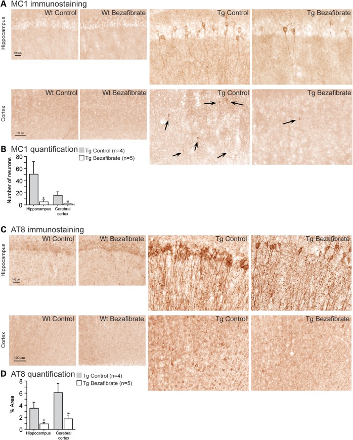
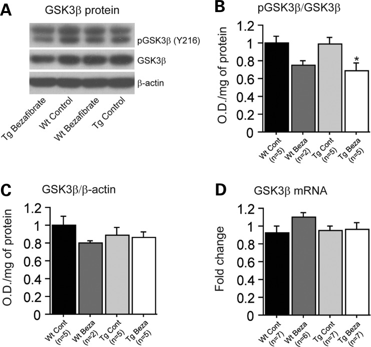
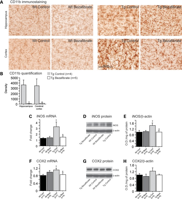
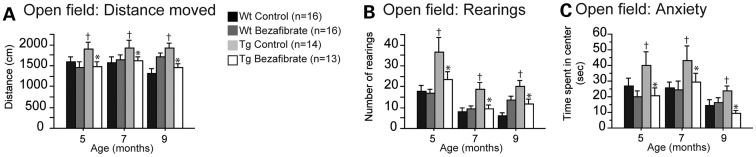
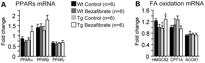
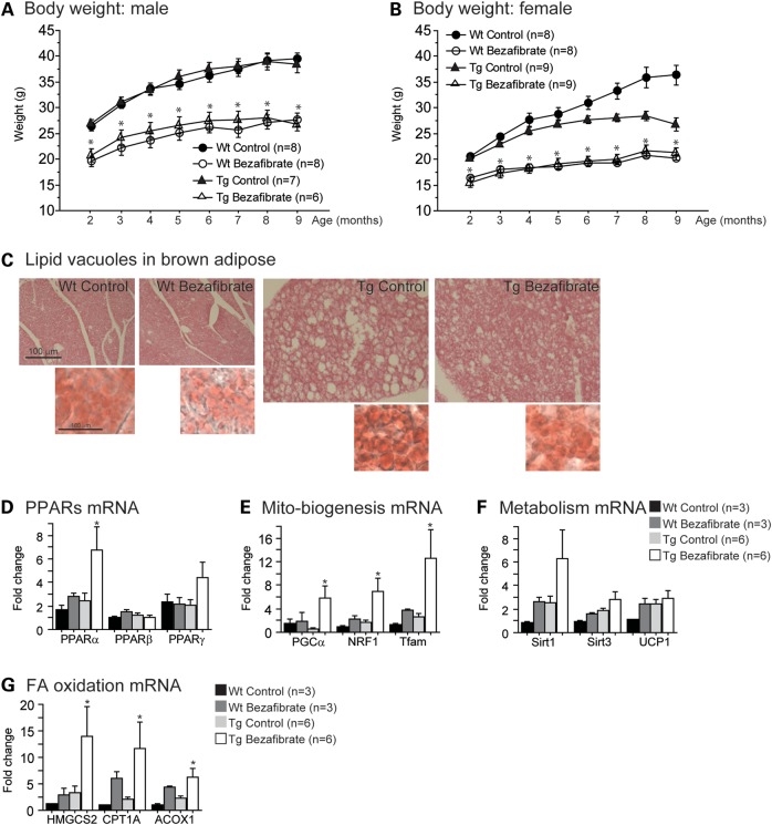
Peroxisome proliferator-activated receptors (PPARs) are ligand-mediated transcription factors, which control both lipid and energy metabolism and inflammation pathways. PPARγ agonists are effective in the treatment of metabolic diseases and, more recently, neurodegenerative diseases, in which they show promising neuroprotective effects. We studied the effects of the pan-PPAR agonist bezafibrate on tau pathology, inflammation, lipid metabolism and behavior in transgenic mice with the P301S human tau mutation, which causes familial frontotemporal lobar degeneration. Bezafibrate treatment significantly decreased tau hyperphosphorylation using AT8 staining and the number of MC1-positive neurons. Bezafibrate treatment also diminished microglial activation and expression of both inducible nitric oxide synthase and cyclooxygenase 2. Additionally, the drug differentially affected the brain and brown fat lipidome of control and P301S mice, preventing lipid vacuoles in brown fat. These effects were associated with behavioral improvement, as evidenced by reduced hyperactivity and disinhibition in the P301S mice. Bezafibrate therefore exerts neuroprotective effects in a mouse model of tauopathy, as shown by decreased tau pathology and behavioral improvement. Since bezafibrate was given to the mice before tau pathology had developed, our data suggest that bezafibrate exerts a preventive effect on both tau pathology and its behavioral consequences. Bezafibrate is therefore a promising agent for the treatment of neurodegenerative diseases associated with tau pathology.


Figures

Figure 1.
Figure 1.
Bezafibrate reduced tau pathology and tau hyperphosphorylation in P301S mice. (A) Immunohistochemical staining with the MC1 antibody in the hippocampus and cerebral cortex (scale bar: 100 μm). (B) Number of MC1-positive neurons in the hippocampus and cerebral cortex of P301S mice fed a control diet (Tg Control, n = 4) and P301S mice fed a bezafibrate diet (Tg Bezafibrate, n = 5). (C) Immunohistochemical staining with the AT8 antibody to phosphorylated tau in the hippocampus and cerebral cortex (scale bar: 100 μm). (D) Percent area covered by AT8 immunoreactivity in the hippocampus and cerebral cortex of P301S mice fed a control diet (Tg Control, n = 4) and P301S mice fed a bezafibrate diet (Tg Bezafibrate, n = 5). Administration of bezafibrate in P301S mice significantly reduced tau pathology and phosphorylation (unpaired t-tests, *P < 0.05).
Figure 2.
Figure 2.
Bezafibrate treatment affected GSK3β phosphorylation in P301S mice. (A) Western blots of phospho-GSK3β and total GSK3β, and (B and C) quantification by optical densities in wild-type mice fed a control diet (Wt Cont, n = 5), wild-type fed a bezafibrate (Wt Beza, n = 2), P301S mice fed a control diet (Tg Cont, n = 5) and P301S mice fed a bezafibrate diet (Tg Beza, n = 5). (C) Levels of total GSK3β mRNA in wild-type mice fed a control diet (Wt Cont, n = 7), wild-type fed a bezafibrate (Wt Beza, n = 6), P301S mice fed a control diet (Tg Cont, n = 7) and P301S mice fed a bezafibrate diet (Tg Beza, n = 7). Bezafibrate treatment affected the phosphorylation of GSK3β levels in P301S mice, without affecting total GSK3β mRNA and protein levels (Fisher's PLSD, *P < 0.05).
Figure 3.
Figure 3.
Bezafibrate treatment reduced inflammation in P301S mice. (A) Immunohistochemical staining with the CD11b antibody in the hippocampus and cerebral cortex. (B) Intensity (density) of CD11b in P301S mice fed a control diet (Tg Control, n = 4) and P301S mice fed a bezafibrate diet (Tg Bezafibrate, n = 5; scale bar: 100 μm). Administration of bezafibrate significantly reduced microglial activation in the brains of P301S mice (unpaired t-tests, *P < 0.05). Levels of iNOS (C) and COX2 (F) mRNA in wild-type mice fed a control diet (Wt Control, n = 7), wild-type mice fed a bezafibrate diet (Wt Bezafibrate, n = 6), P301S mice fed a control diet (Tg Control, n = 7) and P301S mice fed a bezafibrate diet (Tg Bezafibrate, n = 5). Western blots of iNOS (D) and COX2 (G) protein and their quantifications by optical densities normalized to β-actin (E and H) in wild-type mice fed a control diet (Wt Control, n = 4), wild-type mice fed a bezafibrate diet (Wt Bezafibrate, n = 2), P301S mice fed a control diet (Tg Control, n = 5) and P301S mice fed a bezafibrate diet (Tg Bezafibrate, n = 5). P301S mice had significantly elevated inflammation relative to their wild-type littermates (Fisher's PLSD, P < 0.05). After bezafibrate treatment, both iNOS and COX2 mRNA and protein expression were significantly decreased in P301S mice (Fisher's PLSD, *P < 0.05).
Figure 4.
Figure 4.
Bezafibrate treatment improved behavioral deficits in P301S mice. Distance moved (A), rearings (B) and anxiety (time spent in the center) (C) of the open field in wild-type mice fed a control diet (Wt Control, n = 16), wild-type mice fed a bezafibrate diet (Wt Bezafibrate, n = 16), P301S mice fed a control diet (Tg Control, n = 14) and P301S mice fed a bezafibrate diet (Tg Bezafibrate, n = 13). P301S mice were significantly hyperactive and disinhibited in the open-field test when compared with wild-type littermates (Fisher's PLSD, P < 0.05). Bezafibrate treatment significantly reduced behavioral abnormalities in P301S mice (Fisher's PLSD, *P < 0.05).
Figure 5.
Figure 5.
Bezafibrate treatment increased FA metabolism in P301S brains. Expression of PPAR (A) and fatty β-oxidation genes (B) in the brains of wild-type mice fed a control diet (Wt Control, n = 6), wild-type mice fed a bezafibrate diet (Wt Bezafibrate, n = 6), P301S mice fed a control diet (Tg Control, n = 6) and P301S mice fed a bezafibrate diet (Tg Bezafibrate, n = 6). P301S mice had reduced mRNA levels of HMGCS2 (Fisher's PLSD, P < 0.05). After bezafibrate treatment, there was a trend toward an increase in PPARα and PPARβ, and there was a significant increase in HMGCS2 mRNA levels in P301S mouse brains (Fisher's PLSD, *P < 0.05).
Figure 6.
Figure 6.
Bezafibrate treatment prevented lipid vacuoles and activated mitochondrial biogenesis and β-oxidation genes in brown adipose tissue of P301S mice. Body weight of male (A) and female (B) wild-type mice fed a control diet (Wt Control, total n = 16), wild-type mice fed a bezafibrate diet (Wt Bezafibrate, total n = 16), P301S mice fed a control diet (Tg Control, total n = 16) and P301S mice fed a bezafibrate diet (Tg Bezafibrate, total n = 15). Bezafibrate-treated mice had significantly lower body weight than control-treated mice (Fisher's PLSD, *P < 0.05). (C) Hematoxylin–eosin staining of brown adipose tissue in wild-type mice fed a bezafibrate diet (Wt Bezafibrate, n = 5), P301S mice fed a control diet (Tg Control, n = 5) and P301S mice fed a bezafibrate diet (Tg Bezafibrate, n = 5). (C) Oil red O staining in bottom right panels was used to verify the presence of lipids. P301S mice had increased size and number of lipid vacuoles, pathology which was improved after bezafibrate treatment. Expression of PPARs (D) and energy metabolism-related genes (E and F) in wild-type mice fed a control diet (Wt Control, n = 3), wild-type mice fed a bezafibrate diet (Wt Bezafibrate, n = 3), P301S mice fed a control diet (Tg Control, n = 6) and P301S mice fed a bezafibrate diet (Tg Bezafibrate, n = 6). There were significant increases in PPARα, PGC1α, NRF1 and Tfam in the brown adipose of P301S mice after bezafibrate treatment (Fisher's PLSD, *P < 0.05). Also, there was a trend toward an increase in PPARγ and Sirt1. (G) Expression of FA β-oxidation genes in wild-type mice fed a control diet (Wt Control, n = 3), wild-type mice fed a bezafibrate diet (Wt Bezafibrate, n = 3), P301S mice fed a control diet (Tg Control, n = 6) and P301S mice fed a bezafibrate diet (Tg Bezafibrate, n = 6). There was a significant increase in HMGCS2, CPT1A and ACOX1 in the brown adipose of P301S mice after bezafibrate treatment (Fisher's PLSD, *P < 0.05).

References

    1. Bensinger S.J., Tontonoz P. Integration of metabolism and inflammation by lipid-activated nuclear receptors. Nature. 2008;454:470–477. doi:10.1038/nature07202. - DOI - PubMed
    1. Schulman I.G. Nuclear receptors as drug targets for metabolic disease. Adv. Drug Deliv. Rev. 2010;62:1307–1315. doi:10.1016/j.addr.2010.07.002. - DOI - PMC - PubMed
    1. Wang Y.X. PPARs: diverse regulators in energy metabolism and metabolic diseases. Cell Res. 2010;20:124–137. doi:10.1038/cr.2010.13. - DOI - PMC - PubMed
    1. Mandrekar-Colucci S., Landreth G.E. Nuclear receptors as therapeutic targets for Alzheimer's disease. Expert Opin. Ther. Targets. 2011;15:1085–1097. doi:10.1517/14728222.2011.594043. - DOI - PMC - PubMed
    1. Kaundal R.K., Sharma S.S. Peroxisome proliferator-activated receptor gamma agonists as neuroprotective agents. Drug News Perspect. 2010;23:241–256. doi:10.1358/dnp.2010.23.4.1437710. - DOI - PubMed

Publication types

MeSH terms