. 2012 Sep;122(9):3330-42.
doi: 10.1172/JCI63004. Epub 2012 Aug 27.

Mouse fukutin deletion impairs dystroglycan processing and recapitulates muscular dystrophy

Affiliations

Mouse fukutin deletion impairs dystroglycan processing and recapitulates muscular dystrophy

Aaron M Beedle et al. J Clin Invest. 2012 Sep.

Abstract

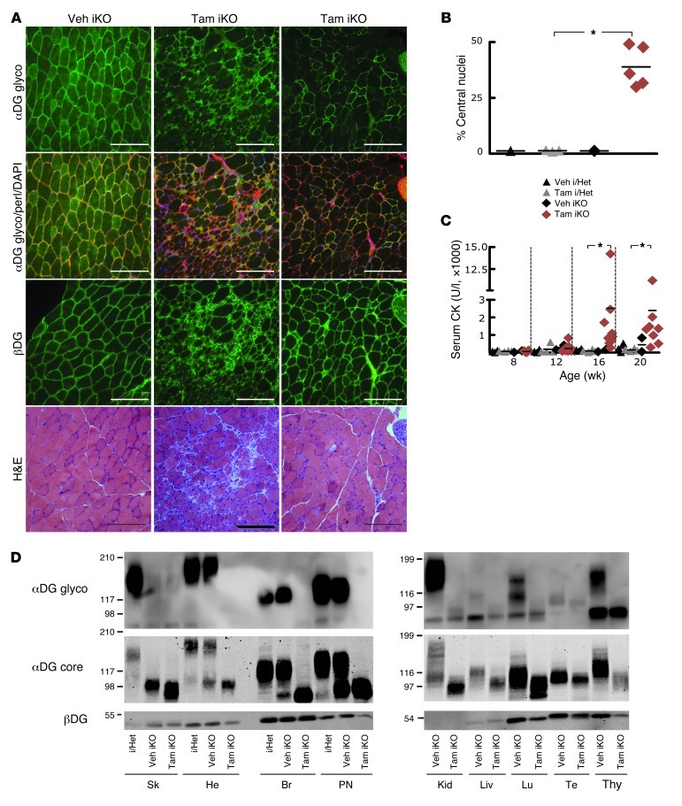
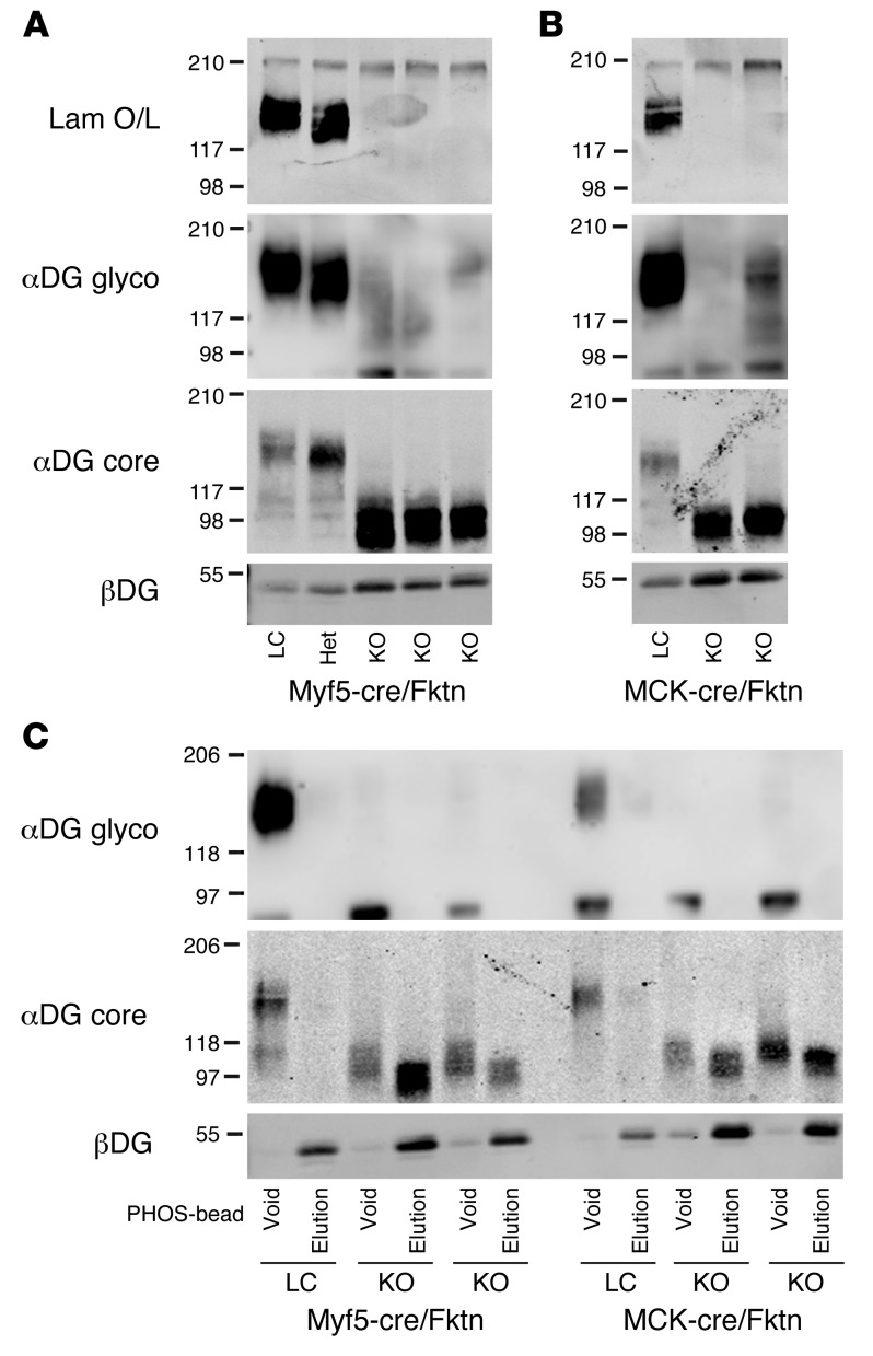
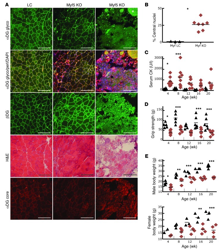
Dystroglycan is a transmembrane glycoprotein that links the extracellular basement membrane to cytoplasmic dystrophin. Disruption of the extensive carbohydrate structure normally present on α-dystroglycan causes an array of congenital and limb girdle muscular dystrophies known as dystroglycanopathies. The essential role of dystroglycan in development has hampered elucidation of the mechanisms underlying dystroglycanopathies. Here, we developed a dystroglycanopathy mouse model using inducible or muscle-specific promoters to conditionally disrupt fukutin (Fktn), a gene required for dystroglycan processing. In conditional Fktn-KO mice, we observed a near absence of functionally glycosylated dystroglycan within 18 days of gene deletion. Twenty-week-old KO mice showed clear dystrophic histopathology and a defect in glycosylation near the dystroglycan O-mannose phosphate, whether onset of Fktn excision driven by muscle-specific promoters occurred at E8 or E17. However, the earlier gene deletion resulted in more severe phenotypes, with a faster onset of damage and weakness, reduced weight and viability, and regenerating fibers of smaller size. The dependence of phenotype severity on the developmental timing of muscle Fktn deletion supports a role for dystroglycan in muscle development or differentiation. Moreover, given that this conditional Fktn-KO mouse allows the generation of tissue- and timing-specific defects in dystroglycan glycosylation, avoids embryonic lethality, and produces a phenotype resembling patient pathology, it is a promising new model for the study of secondary dystroglycanopathy.


Figures

Figure 1
Figure 1. Inducible, whole-animal Fktn deletion causes dystrophic features with disruption of αDG glycosylation.
(A) Representative iliopsoas images from a control TamCre-Esr1/Fktnfl/– mouse (Veh iKO) and 2 treated TamCre-Esr1/Fktnfl/– mice (Tam iKO) at 20 weeks (Tam dosed at 6 weeks of age). Dystrophic pathology is evident in Tam iKO muscle with H&E staining. αDG glycosylation (αDG glyco) is abnormal in KO mice, while βDG is unchanged. ECM protein perlecan (perl) and nuclear (DAPI) counterstains are shown; original magnification, ×20; scale bars: 100 μm. (B) The percentage of centrally nucleated fibers is plotted for individual mice. i/Het, heterozygous or inducible heterozygous. *P = 0.036, Tam i/Het versus Tam iKO; Mann-Whitney test. (C) Serum CK activity is plotted for individual mice at various ages. *P ≤ 0.05; Dunn’s test. (D) DG expression and glycosylation in various tissues. αDG glyco detection by IIH6 is disrupted in all Tam iKO tissues tested, and core αDG protein (αDG core) is reduced in mass. An intermediate phenotype was observed in some Veh iKO mice, indicating exposure to Tam from treated littermates. βDG blots demonstrate different DG expression levels across tissues and confirm the presence of DG in Fktn-deficient mice. Sk, skeletal muscle; He, heart; Br, brain; PN, peripheral nerve; Kid, kidney; Liv, liver; Lu, lung; Te, testes; Thy, thymus.
Figure 2
Figure 2. Biochemical analyses of dystroglycan in skeletal muscle from Fktn-KO mice and littermates.
(A) Western blot analyses of WGA-purified skeletal muscle from myf5-Cre/Fktn (E8) KO mice and littermates (LC, Het). All KOs have reduced αDG mass, as measured by detection of core protein (αDG core). This corresponds to a complete loss of αDG binding activity in laminin overlay (Lam O/L) and loss or reduction of αDG glyco-epitopes (αDG glyco). βDG protein is detected in all cases. (B) Western blot analyses of MCK-Cre/Fktn (E17) KO and littermates, as in A. (C) Detection of αDG free phosphate. WGA-enriched skeletal muscle was applied to PHOS-beads to capture proteins with exposed phosphates. αDG from control mice has no free phosphate, but E8 and E17 muscle KOs (myf5-Cre/Fktn KO, MCK-Cre/Fktn KO) contain a substantial proportion with exposed phosphate. βDG is normally phosphorylated and serves as an experimental control.
Figure 3
Figure 3. Cre recombinase driven at E17 by the MCK promoter directs Fktn deletion in differentiating striated muscle to cause phenotypes consistent with mild muscular dystrophy.
(A) H&E and immunofluorescence images from the iliopsoas of 20-week-old Het and KO mice. Dystrophic pathology is observed in KO mice along with patchy expression of glycosylated αDG (αDG glyco). αDG core protein and βDG are unchanged. DAPI nuclear stain and perlecan are shown for comparison; original magnification, ×20; scale bars: 100 μm. (B) The percentage of iliopsoas fibers with central nucleation is shown for individual 20-week-old mice. ***P = 0.0007; Mann Whitney test. (C) CK activity is detected in serum of individual littermate and KO mice at various ages. **P = 0.001–0.01, 12-week MCK LC versus KO; Bonferroni test. (D) Analysis of average forelimb grip strength for individual mice at various ages did not reveal muscle weakness in KO mice. P > 0.05; ANOVA. (E) Body weights of male and female LC and KO mice from 4 through 20 weeks (P > 0.05; ANOVA). Black triangles, MCK-Cre LC; red diamonds, MCK-Cre Fktn-KO. Statistics were calculated for LC versus KO mice at each age.
Figure 4
Figure 4. Mice with skeletal muscle Fktn deletion initiated at E8 show moderate to severe muscular dystrophy.
(A) Histology of iliopsoas muscle from 20-week-old myf5-Cre LC and KO mice provides evidence of moderate to severe dystrophy. Patchy or absent staining with αDG glyco-antibody while βDG and αDG core protein are still present indicates abnormal αDG glycosylation. Original magnification, ×20; scale bars: 100 μm. Note: Peripheral nerve twigs are still αDG glyco positive, confirming specificity of Fktn KO in muscle (e.g., asterisk). (B) Iliopsoas fibers with central nucleation in individual mice at 20 weeks of age. *P = 0.017; Mann Whitney test. (C) CK activity is elevated in KO mice at young ages. *P = 0.01–0.05, 4-week Myf LC versus KO; ***P < 0.001, 8-week Myf LC versus KO; Bonferroni test. (D) Forelimb grip strength (average of 5 pulls) is plotted according to age (*P = 0.01–0.05, 4-week Myf LC versus KO; ***P < 0.001, 8-, 16-, and 20-week Myf LC versus KO pairs; Bonferroni test. (E) Body weights of male and female mice at various ages. *P = 0.01–0.05, 4-week Myf LC versus KO M; **P = 0.001–0.01, 8-week Myf LC versus KO male and 16-week Myf LC versus KO female; ***P < 0.001, 12-, 16-, and 20-week Myf LC versus KO paired male and 20-week Myf LC versus KO female; Bonferonni test. Each data point represents 1 mouse; group means are shown. Black triangles, myf5-Cre LC; red diamonds, myf5-Cre Fktn-KO. Statistics were calculated for LC versus KO mice at each age.

Comment in

References

    1. Ervasti JM, Campbell KP. Membrane organization of the dystrophin-glycoprotein complex. Cell. 1991;66(6):1121–1131. doi: 10.1016/0092-8674(91)90035-W. - DOI - PubMed
    1. Ervasti JM. Dystrophin, its interactions with other proteins, and implications for muscular dystrophy. Biochim Biophys Acta. 2007;1772(2):108–117. - PubMed
    1. Guglieri M, Magri F, Comi GP. Molecular etiopathogenesis of limb girdle muscular and congenital muscular dystrophies: boundaries and contiguities. Clin Chim Acta. 2005;361(1–2):54–79. doi: 10.1016/j.cccn.2005.05.020. - DOI - PubMed
    1. Ibraghimov-Beskrovnaya O, Ervasti JM, Leveille CJ, Slaughter CA, Sernett SW, Campbell KP. Primary structure of dystrophin-associated glycoproteins linking dystrophin to the extracellular matrix. Nature. 1992;355(6362):696–702. doi: 10.1038/355696a0. - DOI - PubMed
    1. Barresi R, Campbell KP. Dystroglycan: from biosynthesis to pathogenesis of human disease. J Cell Sci. 2006;119(pt 2):199–207. doi: 10.1242/jcs.02814. - DOI - PubMed

Publication types