

Expansion of HIV-specific T follicular helper cells in chronic HIV infection
- PMID: 22922259
- PMCID: PMC3428098
- DOI: 10.1172/JCI64314
Expansion of HIV-specific T follicular helper cells in chronic HIV infection
Abstract
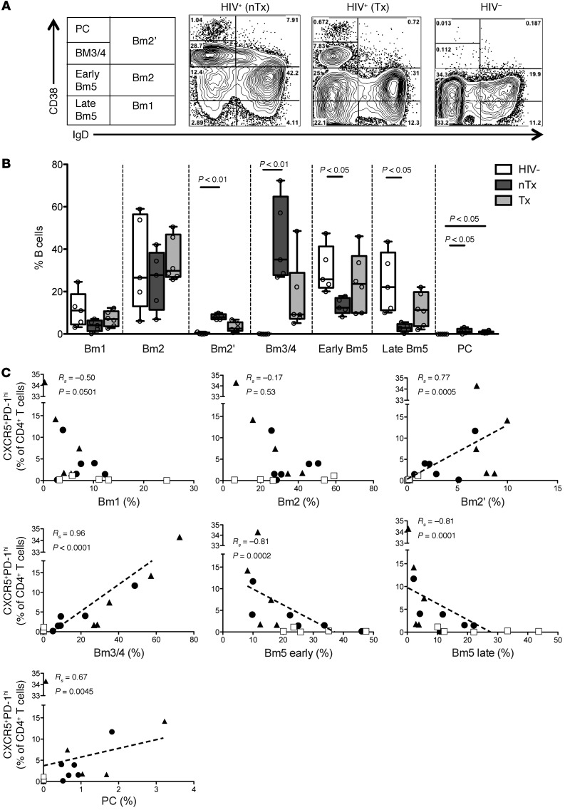
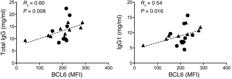
HIV targets CD4 T cells, which are required for the induction of high-affinity antibody responses and the formation of long-lived B cell memory. The depletion of antigen-specific CD4 T cells during HIV infection is therefore believed to impede the development of protective B cell immunity. Although several different HIV-related B cell dysfunctions have been described, the role of CD4 T follicular helper (TFH) cells in HIV infection remains unknown. Here, we assessed HIV-specific TFH responses in the lymph nodes of treatment-naive and antiretroviral-treated HIV-infected individuals. Strikingly, both the bulk TFH and HIV-specific TFH cell populations were significantly expanded in chronic HIV infection and were highly associated with viremia. In particular, GAG-specific TFH cells were detected at significantly higher levels in the lymph nodes compared with those of GP120-specific TFH cells and showed preferential secretion of the helper cytokine IL-21. In addition, TFH cell expansion was associated with an increase of germinal center B cells and plasma cells as well as IgG1 hypersecretion. Thus, our study suggests that high levels of HIV viremia drive the expansion of TFH cells, which in turn leads to perturbations of B cell differentiation, resulting in dysregulated antibody production.
Figures


Comment in
-
HIV and T follicular helper cells: a dangerous relationship.J Clin Invest. 2012 Sep;122(9):3059-62. doi: 10.1172/JCI65175. Epub 2012 Aug 27. J Clin Invest. 2012. PMID: 22922252 Free PMC article.
Similar articles
-
Follicular helper T cells serve as the major CD4 T cell compartment for HIV-1 infection, replication, and production.J Exp Med. 2013 Jan 14;210(1):143-56. doi: 10.1084/jem.20121932. Epub 2012 Dec 17. J Exp Med. 2013. PMID: 23254284 Free PMC article.
-
Activation of CXCR3+ Tfh cells and B cells in lymph nodes during acute HIV-1 infection correlates with HIV-specific antibody development.J Virol. 2025 Mar 18;99(3):e0153224. doi: 10.1128/jvi.01532-24. Epub 2025 Feb 11. J Virol. 2025. PMID: 39932316 Free PMC article.
-
Early T Follicular Helper Cell Responses and Germinal Center Reactions Are Associated with Viremia Control in Immunized Rhesus Macaques.J Virol. 2019 Feb 5;93(4):e01687-18. doi: 10.1128/JVI.01687-18. Print 2019 Feb 15. J Virol. 2019. PMID: 30463978 Free PMC article.
-
Follicular CD4 T Helper Cells As a Major HIV Reservoir Compartment: A Molecular Perspective.Front Immunol. 2018 Jun 18;9:895. doi: 10.3389/fimmu.2018.00895. eCollection 2018. Front Immunol. 2018. PMID: 29967602 Free PMC article. Review.
-
Interleukin-21 and T follicular helper cells in HIV infection: research focus and future perspectives.Immunol Res. 2013 Dec;57(1-3):279-91. doi: 10.1007/s12026-013-8457-0. Immunol Res. 2013. PMID: 24242760 Free PMC article. Review.
Cited by
-
A Blueprint for HIV Vaccine Discovery.Cell Host Microbe. 2012 Oct 18;12(4):396-407. doi: 10.1016/j.chom.2012.09.008. Cell Host Microbe. 2012. PMID: 23084910 Free PMC article. Review.
-
Insights into the development and regulation of T follicular helper cells.Cytokine. 2016 Nov;87:9-19. doi: 10.1016/j.cyto.2016.06.010. Epub 2016 Jun 20. Cytokine. 2016. PMID: 27339151 Free PMC article. Review.
-
The Role of Immunomodulatory Receptors in the Pathogenesis of HIV Infection: A Therapeutic Opportunity for HIV Cure?Front Immunol. 2020 Jul 2;11:1223. doi: 10.3389/fimmu.2020.01223. eCollection 2020. Front Immunol. 2020. PMID: 32714317 Free PMC article. Review.
-
Subversion of the B-cell compartment during parasitic, bacterial, and viral infections.BMC Immunol. 2015 Mar 26;16:15. doi: 10.1186/s12865-015-0079-y. BMC Immunol. 2015. PMID: 25884828 Free PMC article. Review.
-
Control of the Germinal Center by Follicular Regulatory T Cells During Infection.Front Immunol. 2018 Nov 20;9:2704. doi: 10.3389/fimmu.2018.02704. eCollection 2018. Front Immunol. 2018. PMID: 30524440 Free PMC article. Review.
References
Publication types
MeSH terms
Substances
Grants and funding
LinkOut - more resources
Full Text Sources
Other Literature Sources
Medical
Research Materials
