. 2012 Sep;122(9):3145-58.
doi: 10.1172/JCI63401. Epub 2012 Aug 27.

Autonomic neurocristopathy-associated mutations in PHOX2B dysregulate Sox10 expression

Affiliations

Autonomic neurocristopathy-associated mutations in PHOX2B dysregulate Sox10 expression

Mayumi Nagashimada et al. J Clin Invest. 2012 Sep.

Abstract

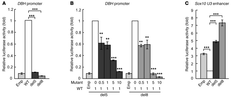
The most common forms of neurocristopathy in the autonomic nervous system are Hirschsprung disease (HSCR), resulting in congenital loss of enteric ganglia, and neuroblastoma (NB), childhood tumors originating from the sympathetic ganglia and adrenal medulla. The risk for these diseases dramatically increases in patients with congenital central hypoventilation syndrome (CCHS) harboring a nonpolyalanine repeat expansion mutation of the Paired-like homeobox 2b (PHOX2B) gene, but the molecular mechanism of pathogenesis remains unknown. We found that introducing nonpolyalanine repeat expansion mutation of the PHOX2B into the mouse Phox2b locus recapitulates the clinical features of the CCHS associated with HSCR and NB. In mutant embryos, enteric and sympathetic ganglion progenitors showed sustained sex-determining region Y (SRY) box10 (Sox10) expression, with impaired proliferation and biased differentiation toward the glial lineage. Nonpolyalanine repeat expansion mutation of PHOX2B reduced transactivation of wild-type PHOX2B on its known target, dopamine β-hydroxylase (DBH), in a dominant-negative fashion. Moreover, the introduced mutation converted the transcriptional effect of PHOX2B on a Sox10 enhancer from repression to transactivation. Collectively, these data reveal that nonpolyalanine repeat expansion mutation of PHOX2B is both a dominant-negative and gain-of-function mutation. Our results also demonstrate that Sox10 regulation by PHOX2B is pivotal for the development and pathogenesis of the autonomic ganglia.


Figures

Figure 1
Figure 1. Generation of NPARM PHOX2B knockin mice.
(A) Protein structure of WT and NPARM (931 del5 and 693–700 del8) PHOX2B. Red arrows indicate the regions of the deletions. Dark gray boxes depict aberrant 42 amino acids shared by both del5 and del8 mutant proteins. HD, homeodomain; 9, 20, polyalanine stretches. (B) Schematics showing the gene-targeting strategy. Gray boxes indicate exons of the Phox2b gene. Red boxes show human genomic fragments spanning from the deletion mutations to the aberrant stop codon due to the shift in the ORF. A floxed (white triangles) Tn5-neo cassette was removed in vivo by Cre recombination. Bars depict the location of the probe for Southern blot analysis and PCR primers for screening the recombined ES clones. (C) Southern blot and PCR analysis of WT and the targeted allele of the Phox2b gene. Properly targeted ES clones (Mut) display both WT (14.5 kb) and mutant (7 kb) fragments. (D) ISH analysis of embryonic gut (E13.5). NPARM PHOX2B–specific riboprobes detected signals in enteric ganglion progenitors of del8 mutant but not in WT gut (bottom panels). Anti-WT Phox2b antibodies were used to visualize enteric ganglion progenitors (top panels). Scale bar: 50 μm.
Figure 2
Figure 2. NPARM PHOX2B mutant mice display phenotypes reminiscent of CCHS-HSCR-NB association.
(A) WT (top 4) and del8 mutant (bottom 4) mice born by Caesarian section at E18.5. Note cyanotic skin color of the mutant mice. (B) Schematic representation of various brain nuclei in a parasagittal section of the hindbrain. nVII, facial motor nucleus; BC, Bötzinger complex; pBC, pre-Bötzinger complex; VRG, ventral respiratory group. (C) Phox2b immunostaining (top) of the hindbrain sections (E18.5) and Nissl staining of the adjacent sections (bottom). Structure of both nVII (arrows) and RTN/pFRG (arrowheads in WT) is severely disrupted in NPARM PHOX2B mutant mice. (D) Whole-mount acetylcholine esterase staining of the gut. Note the oligoganglionosis and aganglionosis of the colon in del5 and del8 mutant mice, respectively. Thick nerve bundles seen in the colon of del8 mutants are extrinsic nerve fibers that failed to defasciculate due to the absence of the enteric ganglia (61). (E and F) Whole mount TH staining of the thorax (E) and pararenal areas (F). Arrowheads indicate aberrantly located ganglia. Note that individual ganglia of the thoracic chain (dotted circles in bottom panels of E) of the mutants are smaller in size and harbor thinner nerve fibers than those of WT. Scale bars: 100 μm (C, D [intestine and colon]); 330 μm (E [bottom panels]); 1 mm (D [stomach], E [top panels], F).
Figure 3
Figure 3. Impaired migration and differentiation of enteric ganglion progenitors in NPARM PHOX2B embryos.
(AC) ISH analysis of Sox10 expression in ENCCs. Arrows indicate the foreguts of E10.5 embryos. Insets in the upper-right corners are larger magnifications of boxed areas. (DF and HJ) Phox2b/Sox10 double immunostaining of E12 gut. Invasion of ENCCs in the hindgut mesenchyme (D) is retarded in NPARM PHOX2B embryos (E and F). ENCC density was noticeably lower in the mutant than in WT midgut. (G) Schematic representation showing the gut morphology at E12. Areas covered by ENCCs in WT hindgut are shown in green. (HJ) Larger magnification of the midgut regions. Arrows indicate neuronally committed cells expressing WT Phox2b, but not Sox10. (KP) TuJ1 staining of embryonic gut. TuJ1+ cells are drastically decreased in NPARM PHOX2B embryos (L and M). Extinguishment of Sox10 expression in these early differentiating neurons is also impaired in NPARM PHOX2B embryos (arrows in O and P). Scale bars: 10 μm (P); 20 μm (J); 200 μm (C, F, and M).
Figure 4
Figure 4. Abnormal neuroglial differentiation of sympathetic ganglion cells in NPARM PHOX2B embryos.
(A) Expression of WT Phox2b and Sox10 in the nascent sympathetic ganglia. Note that Sox10+Phox2b cells (white arrows) are more prominent in NPARM PHOX2B sympathetic ganglia. (B) Expression of various markers in the sympathetic chain. First and second rows represent TH staining of whole-mount preparations and transverse sections of E10.5 embryos. The third through sixth rows show results of ISH analysis. (C and E) Analysis of neuroglial differentiation in developing sympathetic ganglia by Phox2b/Sox10 immunohistochemistry. Although Sox10+ cells (satellite cell progenitors) are localized preferentially at the periphery of the WT ganglia at E11.5 (C, white arrowheads), many Sox10+ cells are also found inside of the sympathetic ganglia in NPARM PHOX2B embryos (C, arrows). (D) BrdU (red) and WT Phox2b (green) double labeling of E11.5 sympathetic chain ganglia. (E) Note that, at E18.5, Sox10+ cells appear more prominent in NPARM PHOX2B than in WT sympathetic ganglia (SCG). Scale bars: 20 μm (A, CE); 150 μm (B, TH, Dbh, Ret, Ascl1, Erbb3); 500 μm (B, TH, whole-mount).
Figure 5
Figure 5. Characterization of the sympathetic ganglion progenitors by a neurosphere method.
(A and B) Morphology and diameter of the primary neurospheres generated from dissociated sympathetic ganglia (E13.5). (C) A graph showing neurosphere-forming ability. The numbers of neurospheres generated from 5,000 cells is shown as frequency (%). (D) Representative images for Phox2b/Sox10 expression in neurosphere cells cultured on monolayer (passage 1). The vast majority of the NPARM PHOX2B mutant–derived neurosphere cells express both Phox2b and Sox10 (bottom panels). Note also that Phox2b+Sox10 cells are very few in the NPARM PHOX2B mutant–derived neurosphere cells (white arrows). (E) Immunocytochemical analysis of differentiating neurons (passage 1). TuJ1+ cells derived from NPARM PHOX2B neurosphere cells bear thick neurites and aberrantly express Sox10 (white arrows in bottom panels). Scale bars: 20 μm (E); 40 μm (D); 200 μm (A). Error bars indicate SD (n = 3). ***P < 0.001.
Figure 6
Figure 6. Transcriptional properties of WT and NPARM PHOX2B on DBH and Sox10 U3 reporter genes.
Transcriptional activation analysis was performed in NIH3T3 cells using WT and NPARM PHOX2B expression constructs along with DBH or Sox10 U3 reporter genes. (A) Transactivation of the DBH reporter gene. (B) Interaction between WT and NPARM PHOX2B on the DBH reporter activation. Transactivation of the DBH with a constant amount of WT PHOX2B expression construct and increasing amounts of NPARM PHOX2B expression constructs (from 1:0 to 1:10). (C) Effects of WT and NPARM PHOX2B on U3 enhancer of the Sox10 gene. Error bars indicate SD (n = 3). **P < 0.01; ***P < 0.001.
Figure 7
Figure 7. Schematic showing mechanisms underlying autonomic neurocristopathy by NPARM PHOX2B.
Regulation Phox2b and Sox10 expression by reciprocal suppression between these 2 transcription factors plays an important role in generating appropriate numbers of neurons and glia in the enteric and sympathetic ganglia (left). NPARM PHOX2B impairs Phox2b’s inhibitory activity on Sox10 expression and simultaneously transactivates the Sox10 gene, leading to biased differentiation of bipotential progenitors toward glial lineage.

Comment in

Similar articles

Cited by

References

    1. Le Douarin NM. Cell line segregation during peripheral nervous system ontogeny. Science. 1986;231(4745):1515–1522. doi: 10.1126/science.3952494. - DOI - PubMed
    1. Le Douarin NM, Kalcheim C. The Neural Crest. Cambridge, United Kingdom: Cambridge University Press; 1999.
    1. Bolande RP. The neurocristopathies: A unifying concept of disease arising in neural crest. Hum Pathol. 1974;5:409–429. doi: 10.1016/S0046-8177(74)80021-3. - DOI - PubMed
    1. Bolande RP. Neurocristopathy: its growth and development in 20 years. Pediatr Pathol Lab Med. 1997;17(1):1–25. doi: 10.1080/107710497174994. - DOI - PubMed
    1. Etchevers HC, Amiel J, Lyonnet S. Neural Crest Induction and Differentiation. 2006. Molecular bases of human neurocristopathies. In: Saint-Jeannet JP, ed. pp. 213–234. Georgetown, Texas, USA: Landes Bioscience; - PubMed

Publication types

MeSH terms

Supplementary concepts