. 2012 Sep;30(9):876-82.
doi: 10.1038/nbt.2328.

Directed differentiation of human pluripotent stem cells into mature airway epithelia expressing functional CFTR protein

Affiliations

Directed differentiation of human pluripotent stem cells into mature airway epithelia expressing functional CFTR protein

Amy P Wong et al. Nat Biotechnol. 2012 Sep.

Abstract

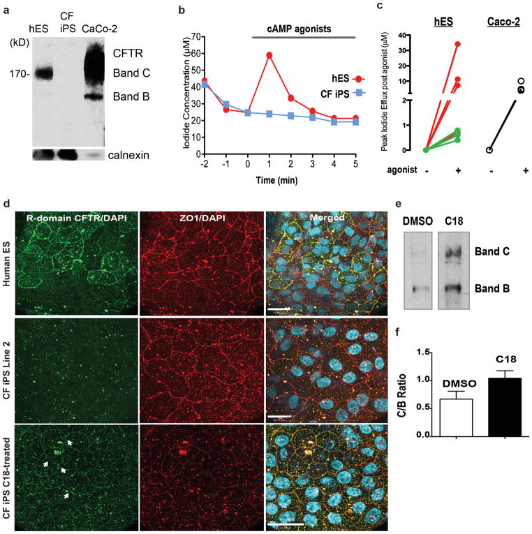
Cystic fibrosis (CF) is a fatal genetic disease caused by mutations in the CFTR (cystic fibrosis transmembrane conductance regulator) gene, which regulates chloride and water transport across all epithelia and affects multiple organs, including the lungs. Here we report an in vitro directed differentiation protocol for generating functional CFTR-expressing airway epithelia from human embryonic stem cells. Carefully timed treatment by exogenous growth factors that mimic endoderm developmental pathways in vivo followed by air-liquid interface culture results in maturation of patches of tight junction–coupled differentiated airway epithelial cells that demonstrate active CFTR transport function. As a proof of concept, treatment of CF patient induced pluripotent stem cell–derived epithelial cells with a small-molecule compound to correct for the common CF processing mutation resulted in enhanced plasma membrane localization of mature CFTR protein. Our study provides a method for generating patient-specific airway epithelial cells for disease modeling and in vitro drug testing.


Conflict of interest statement

Competing Interests Statement

The authors declare that they have no competing financial interests.

Figures

Figure 1
Figure 1. Low concentration of BMP4 up-regulates genes associated with early proximal lung progenitors
a–b. Differentiation to immature lung endoderm cells. c. Expression of airway genes such as KRT5 and Trp63 (basal cell marker), FOXJ1 and SOX17 (ciliated cell markers), NKX2.1 (earliest marker of the lung endoderm), CFTR and the pan-endoderm marker FOXA2 were up-regulated with 10ng/ml of BMP4 (treatment D). Other airway marker MUC5AC (goblet cell marker) was not detected. In addition, the Type II alveolar cell marker surfactant protein-C (SFTPC) and the Clara cell marker SCGB1A1 were not detected up-regulated. SOX9, a marker of the distal tip progenitors found in the developing embryonic lung was also detected. Genes were normalized to the housekeeping gene β-ACTIN and expressed relative to adult tissue positive control RNA. Error bars are s.e.m (n=3 experiments).
Figure 2
Figure 2. FGF18 promotes proximal airway epithelia formation
a–b. Differentiation to proximal lung cells. c. Expression of proximal lung cell genes KRT5, Trp63, FOXJ1, SOX17, MUC5, CFTR, were up-regulated with 10ng/ml of FGF18 (treatment H). Lower levels of the Clara cell marker SCGB1A1, and no significant detection of SFTPC were detected. Other endoderm lineage markers TG and PAX9, AFP, PDX1 and NKX6.1 were not up-regulated. The transcription factors NKX2.1 and FOXA2 that regulate SCGB1A1 and SFTPC expression as well as the distal tip progenitor marker SOX9 were down regulated (compared to B). Other endoderm lineage markers AFP (liver), PDX1 (pancreas), TG (thyroid) and PAX9 (pharyngeal endoderm) were not detected. d. Representative flow histograms of cells from treatment H reveal approximately one-third of the cells are epithelial (panKRT+), CFTR+, FOXJ1+ and NKX2.1+. A majority of the cells express Trp63 (a homolog of p53 that initiates the stratified program in epithelial cells). Grey solid histograms represent respective isotype controls. e. Representative histograms of BrdU incorporation at different stages of differentiation. f. Average cell proliferation at different stages of differentiation from 4 independent lines. Genes were normalized to the housekeeping gene β-ACTIN and expressed relative to adult lung positive control RNA. Error bars are s.e.m (n=3 experiments). *P<0.01, **P<0.001, ***P<0.05 compared to treatment B.
Figure 3
Figure 3. Air liquid interface induces airway epithelial cell differentiation and promotes apical expression of CFTR
a. Schematic of differentiation protocol to generate mature airway epithelium using air liquid interface to induce polarization and apical expression of CFTR as observed in mature airway epithelium. b. The percentage of CFTR+, panKRT+ and FOXJ1+ cells was higher after 3 weeks of ALI while the number of Trp63+ cells was dramatically reduced. c. Gene expression levels of airway markers FOXJ1, MUC5AC, KRT5, Trp63 and SOX17 were significantly higher or comparable to adult lung tissue. Noticeably, CFTR was also up-regulated. The Clara cell marker SCGB1A1 and Type II alveolar cell marker SFTPC were not significantly up-regulated. Genes were normalized to β-ACTIN and expressed relative to adult lung tissue positive control RNA. Error bars are s.e.m (n=3). *P<0.01, **P<0.001 compared to adult lung tissue. d. To confirm the cells are epithelial, co-staining for ZO1 and pan-cytokeratin marker (panKRT) confirmed co-localization of the two proteins suggesting establishment of a tight epithelium. e. Maximal intensity projections of Z-stack confocal images of a 5 week-old ALI culture of human ESC CA1 line-derived epithelia co-express the tight junction associated protein ZO1 and CFTR (clone L12B4). f. The X-Z planar view of the epithelium show apical localization of the CFTR protein. g. Hematoxylin and eosin staining show cilia on some cells. h. Higher magnification shows ciliated cells (green) and apical localization of CFTR (orange, white arrowheads point to apical CFTR). White bar represents 21 microns. i. Low magnification of an ALI transwell cross-section stained for cilia (beta IV tubulin, green) and CFTR (orange) show non-uniform growth of cells with areas of pseudostratified cells and areas of sparse monolayer cells. White bar represents 90 microns. j. High magnification of a culture stained for MUC5AC (green) on the surface of the cells (arrowheads). White bar represents 60 microns.
Figure 4
Figure 4. Establishment of functional CFTR hESC-derived airway epithelia and correction of CF phenotype in CF-iPSC-derived epithelial cells with a small molecule compound C18
a. Western blot shows Band C appearing (~170kDa) in 6-week old hESC-derived epithelial cells (the CA1 cell line) indicative of the complex glycosylated functional form of CFTR. As a positive control, Caco2, a human colorectal adenocarcinoma epithelial cell line that expresses endogenous CFTR was used. The antibody used in this Western (mAb #660) recognizes a peptides within NBD1 of CFTR). Calnexin was probed for loading control. An iodide efflux assay was performed to assess CFTR function in hESC-derived epithelial cells. The cells were preloaded with NaI and regulated CFTR channel activity was stimulated with the agonists of cyclic AMP including: forskolin (10 μM), isobutylmethyl xanthine (100 μM) and genistein (50 μM) (or VX-532 at 10 μM) to activate and potentiate CFTR channel open time at the cell surface. Iodide flux across the apical side of epithelial cells in ALI cultures reports CFTR channel activity since CFTR is permeable to iodide. Iodide efflux was measured using a iodide sensitive microelectrode. CFTR channel activity was detected as a change in iodide flux one to two minutes after addition of cAMP agonists in certain hESC cultures. b. Representative iodide flux graph shows cyclic AMP agonist induced CFTR activity in a differentiated hESC-derived 6-week old ALI culture but not in CF-iPSC-derived culture. c. The response to stimulation of CFTR in 2 hES cell lines, H9 (red) and CA1 (green) and control Caco-2 cell that line that expresses wild-type CFTR (black, 3 cultures). Each line represents a different responsive culture (i.e., showing an increase in efflux rate within 1–2- minutes of stimulation). Four H9 cultures were responsive from a total of 13 cultures and three CA1 cultures were responsive of 16 studied. The H9 cell line could be differentiated to exhibit relatively robust responses. d. Representative photomicrographs of hESC (CA1 line), CF-iPSC GM00997 Line 2 treated with either DMSO (control) or C18 (10μM) and co-stained for tight junction-associated protein ZO1 and CFTR. This antibody recognizes an epitope in the R-domain. Plasma membrane localization of CFTR was observed (white arrowheads) after 2 days of treatment with C18 in the CF-iPSC-derived epithelial cells but not in DMSO controls. White bar indicates scale in microns. e. Cropped western blot shows the accumulation of Band C (mature complex glycosylated form) in C18-corrected cells while the predominant form of the mutant protein in uncorrected cells is Band B (core-glycosylated, ER-retained protein). The antibody used in this Western (mAb #450) recognizes the CFTR peptide: 698-705). Full length blots are presented in Supplementary Figure 14. We observed a trend towards an increase in the C (complex glycosylated) to B ratio of F508del-CFTR protein in C18 treated cultures (n=4) versus DMSO treated cultures (n=3, f.)

References

    1. Van Haute L, De Block G, Liebaers I, Sermon K, De Rycke M. Generation of lung epithelial-like tissue from human embryonic stem cells. Respir Res. 2009;10:105. - PMC - PubMed
    1. Samadikuchaksaraei A, et al. Derivation of distal airway epithelium from human embryonic stem cells. Tissue Eng. 2006;12:867–875. - PubMed
    1. Wang D, Haviland DL, Burns AR, Zsigmond E, Wetsel RA. A pure population of lung alveolar epithelial type II cells derived from human embryonic stem cells. Proc Natl Acad Sci U S A. 2007;104:4449–4454. - PMC - PubMed
    1. Mou H, et al. Generation of Multipotent Lung and Airway Progenitors from Mouse ESCs and Patient-Specific Cystic Fibrosis iPSCs. Cell Stem Cell. 2012;10:385–397. - PMC - PubMed
    1. Kimura J, Deutsch GH. Key mechanisms of early lung development. Pediatr Dev Pathol. 2007;10:335–347. - PubMed

Publication types

MeSH terms

Substances