. 2012 Sep;14(9):950-7.
doi: 10.1038/ncb2560. Epub 2012 Aug 26.

The BBSome controls IFT assembly and turnaround in cilia

Affiliations

The BBSome controls IFT assembly and turnaround in cilia

Qing Wei et al. Nat Cell Biol. 2012 Sep.

Abstract

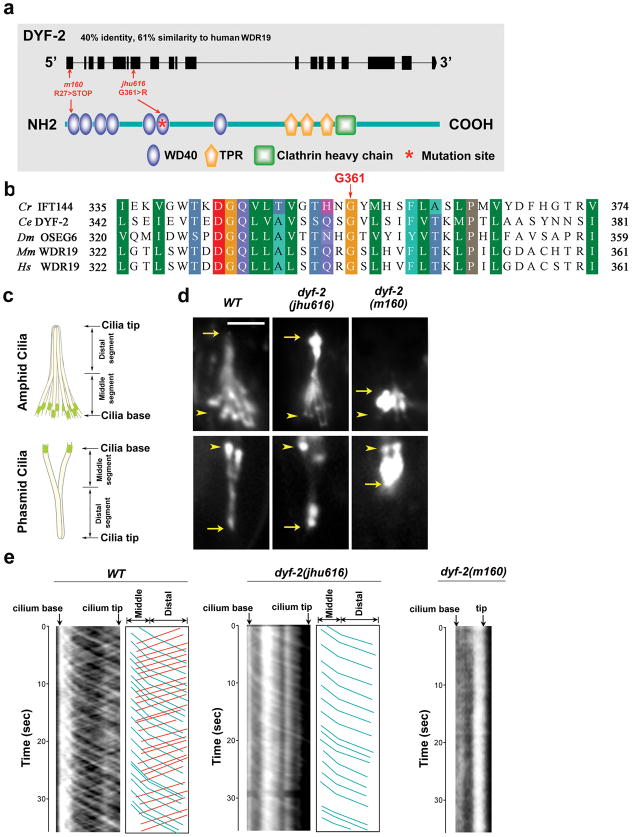
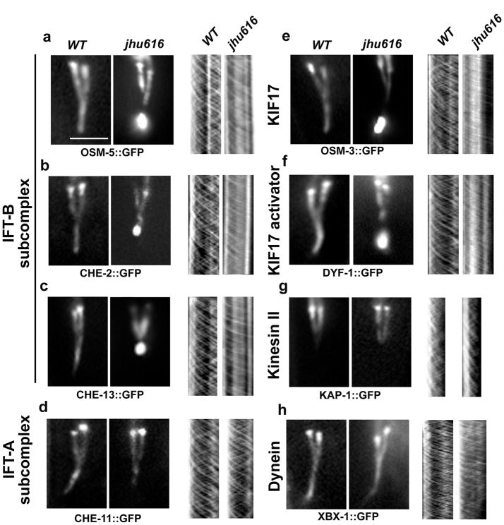
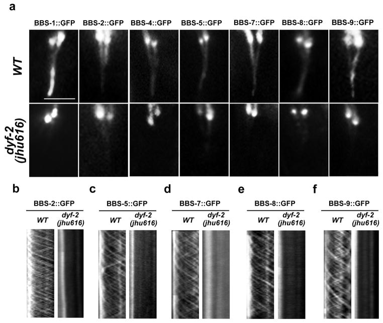
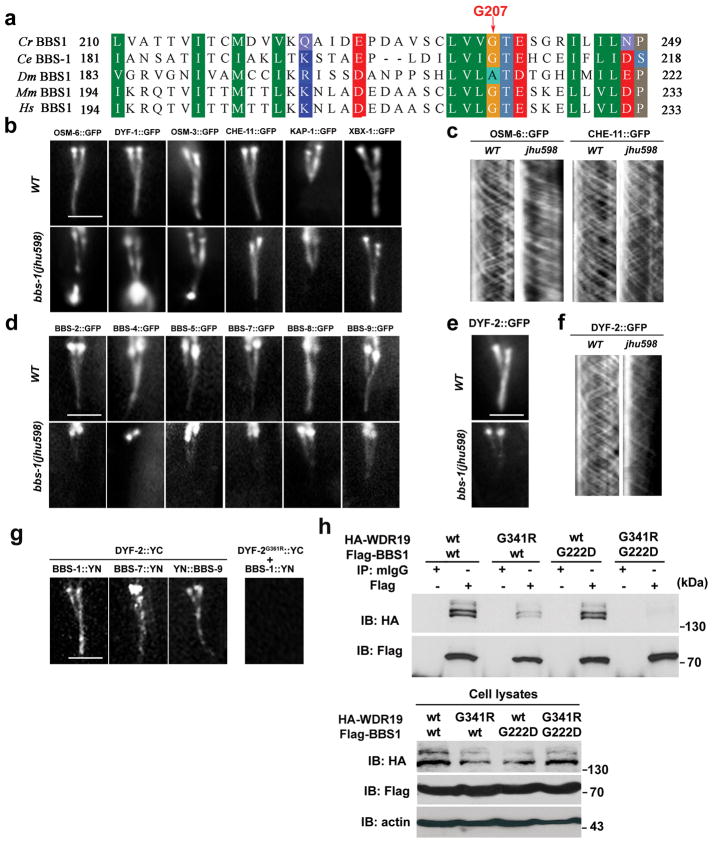
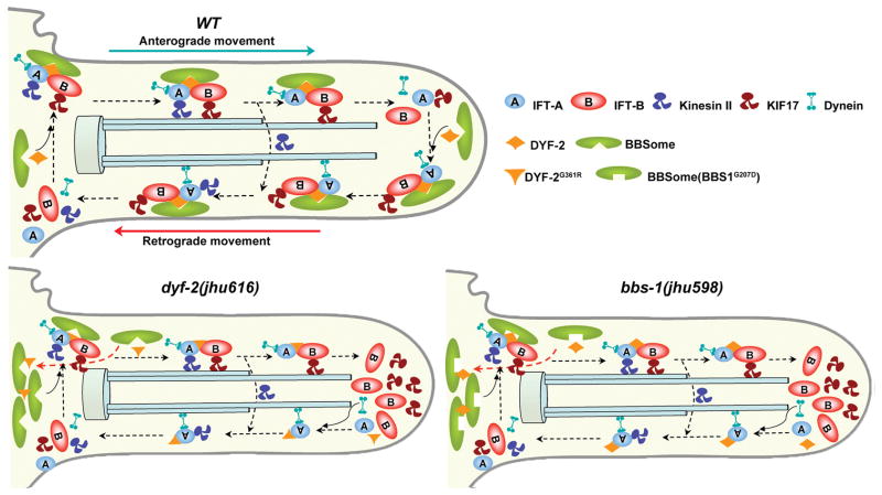
The bidirectional movement of intraflagellar transport (IFT) particles, which are composed of motors, IFT-A and IFT-B subcomplexes, and cargoes, is required for the biogenesis and signalling of cilia(1,2). A successful IFT cycle depends on the proper assembly of the massive IFT particle at the ciliary base and its turnaround from anterograde to retrograde transport at the ciliary tip. However, how IFT assembly and turnaround are regulated in vivo remains elusive. From a whole-genome mutagenesis screen in Caenorhabditis elegans, we identified two hypomorphic mutations in dyf-2 and bbs-1 as the only mutants showing normal anterograde IFT transport but defective IFT turnaround at the ciliary tip. Further analyses revealed that the BBSome (refs 3, 4), a group of conserved proteins affected in human Bardet-Biedl syndrome(5) (BBS), assembles IFT complexes at the ciliary base, then binds to the anterograde IFT particle in a DYF-2- (an orthologue of human WDR19) and BBS-1-dependent manner, and lastly reaches the ciliary tip to regulate proper IFT recycling. Our results identify the BBSome as the key player regulating IFT assembly and turnaround in cilia.


Figures

Figure 1
Figure 1. G361R mutation in the conserved WD40 domain of DYF-2 protein abolishes retrograde IFT transport of OSM-6
a, Schematic of DYF-2 protein and mutation sites. m160 null allele possesses a nonsense mutation at the first exon. b, DYF-2 G361 is highly conserved across species. Cr, Chlamydomonas reinhardtii; Ce, Caenorhabditis elegans; Dm, Drosophila melanogaster; Mm, Mus musculus; Hs, Homo sapiens. c, In C. elegans, amphids in the head and phasmids in the tail are the primary ciliated sensory organs. Amphid and phasmid ciliary axonemes consist of a doublet ‘middle segment’ and a singlet ‘distal segment.’ Amphid channel cilia consist of 10 cilia, and phasmid channel cilia consist of 2 cilia. d, Fluorescence micrographs of cilia labeled with OSM-6::GFP. Compared to the severely truncated cilia in dyf-2(m160) animals, cilia in dyf-2(jhu616) are almost normal. OSM-6::GFP strongly accumulates at the ciliary tip in dyf-2(jhu616). Arrows and arrowheads indicate the tips and bases of cilia, respectively. Bar, 5 μm. e, Kymograph analyses of OSM-6::GFP movement in wild-type, dyf-2(jhu616), and dyf-2(m160) cilia. Kymographs (left panels) and corresponding cartoons (right panels) show visible OSM-6 anterograde (blue lines) and retrograde (red lines) IFT motilities. Compared to wild-type controls, the retrograde transport of OSM-6::GFP is severely abrogated in dyf-2(jhu616) cilia, whereas no IFT movement can be detected in dyf-2(m160) null mutants.
Figure 2
Figure 2. dyf-2(jhu616) animals show compromised IFT turnaround at the ciliary tip
Fluorescence micrographs of cilia labeled with various IFT markers (a–h, left panels) and corresponding kymographs (a–h, right panels). In dyf-2(jhu616), IFT-B components OSM-5 (the ortholog of human IFT88) (a), CHE-2 (the ortholog of human IFT80) (b), and CHE-13 (the ortholog of human IFT57) (c), the IFT-B–associated kinesin motor OSM-3/KIF17 (e), and the OSM-3 activator DYF-1(the homolog of human TTC30B) (f) show consistent ciliary tip accumulation similar to OSM-6 (Fig. 1d) and exhibit severely abrogated retrograde IFT movement (a–c, e–f, right panels). In contrast, IFT-A components CHE-11 (the ortholog of human IFT140) (d) and the IFT-A–associated kinesin-II subunit KAP-1 (g) show no ciliary accumulation and exhibit both anterograde and retrograde IFT motilities at characteristic wild-type rates (Table S1). Note that kinesin-II only mediates IFT motility in the middle segments (g). As expected, the dynein light chain XBX-1 that is responsible for retrograde IFT movement shows no ciliary accumulation and moves normally in both directions (h). Bar, 5 μm.
Figure 3
Figure 3. Lack of functional BBSome in dyf-2(jhu616) cilia
a, BBS proteins show either absent (BBS-1, BBS-4) or dramatically reduced (BBS-2, -5, -7, -8, and -9) ciliary localization in dyf-2(jhu616) animals. b–f, No IFT movements are detected for residual ciliary BBS-2, BBS-5, BBS-7, BBS-8, or BBS-9 in dyf-2(jhu616) cilia. In all panels: bar, 5 μm.
Figure 4
Figure 4. The BBSome and DYF-2 coordinate IFT assembly and turnaround at the ciliary tip
a, BBS-1 G207 site is conserved across species. Cr, Chlamydomonas reinhardtii; Ce, Caenorhabditis elegans; Dm, Drosophila melanogaster; Mm, Mus musculus; Hs, Homo sapiens. b, In bbs-1(jhu598), IFT-B components (OSM-6, DYF-1) and IFT-B associated motor OSM-3, but not IFT-A component CHE-11 and IFT-A associated kinesin-II KAP-1and the retrograde dynein motor XBX-1, accumulate at the cilia tip. c, In bbs-1(jhu598) cilia, the IFT-B protein OSM-6::GFP shows normal anterograde but greatly reduced retrograde movement, whereas the IFT-A protein CHE-11 exhibits normal anterograde and retrograde movements. d, In bbs-1(jhu598), all BBSome subunits show absent or greatly reduced ciliary targeting. e, DYF-2 protein ciliary targeting is also greatly reduced in bbs-1(jhu598) cilia. f, Residual ciliary DYF-2 protein show active bidirectional IFT movement. g, In BiFC analyses, fluorescence complementation can be visualized between YC-tagged DYF-2 and either one of YN-tagged BBS-1, BBS-7, and BBS-9, but not between YC-tagged DYF-2G361R and YN-tagged BBS-1. h, HEK293 cells were transiently transfected with HA-tagged WDR19 and Flag-tagged BBS1 constructs. 48 hours later, cells were subjected to immunoprecipitation using normal mouse IgG (mIgG) or anti-Flag antibody. In all figures: bar, 5 μm.
Figure 5
Figure 5. Model for the role of the BBSome in IFT assembly and turnaround
At the ciliary base in wild-type cells, the BBSome organizes IFT-A, IFT-B, and kinesin motors into a functional complex. DYF-2 stabilizes the association between the BBSome and IFT complex. This stabilization is mediated through the DYF-2 WD40 domain and BBS-1 and may also involve other BBS proteins. IFT particles dissociate after reaching the ciliary tip, and then the BBSome and DYF-2 coordinate to reorganize the entire IFT complex to ready it for retrograde transports. In dyf-2(jhu616) or bbs-1(jhu598) cilia, the BBSome is still functional at the ciliary base, as evident by the observation that IFT-A and IFT-B associate in anterograde transports, whereas the docking of the BBSome onto moving IFT particles is severely abrogated. The absence of the BBSome at the ciliary tip leads to defective reassembly of the IFT complex and the specific accumulation of IFT-B components at the ciliary tip. Note that although DYF-2G361R or DYF-2 in bbs-1(jhu598) is put on moving IFT particles in this model, both of them show severely reduced ciliary targeting.

Comment in

References

    1. Pedersen LB, Rosenbaum JL. Intraflagellar transport (IFT) role in ciliary assembly, resorption and signalling. Current topics in developmental biology. 2008;85:23–61. - PubMed
    1. Scholey JM, Anderson KV. Intraflagellar transport and cilium-based signaling. Cell. 2006;125:439–442. - PubMed
    1. Nachury MV, et al. A core complex of BBS proteins cooperates with the GTPase Rab8 to promote ciliary membrane biogenesis. Cell. 2007;129:1201–1213. - PubMed
    1. Loktev AV, et al. A BBSome subunit links ciliogenesis, microtubule stability, and acetylation. Developmental cell. 2008;15:854–865. - PubMed
    1. Zaghloul NA, Katsanis N. Mechanistic insights into Bardet-Biedl syndrome, a model ciliopathy. The Journal of clinical investigation. 2009;119:428–437. - PMC - PubMed

Publication types

Substances