. 2012 Oct;15(10):1407-13.
doi: 10.1038/nn.3195. Epub 2012 Aug 26.

Lipidomics identifies cardiolipin oxidation as a mitochondrial target for redox therapy of brain injury

Affiliations

Lipidomics identifies cardiolipin oxidation as a mitochondrial target for redox therapy of brain injury

Jing Ji et al. Nat Neurosci. 2012 Oct.

Abstract

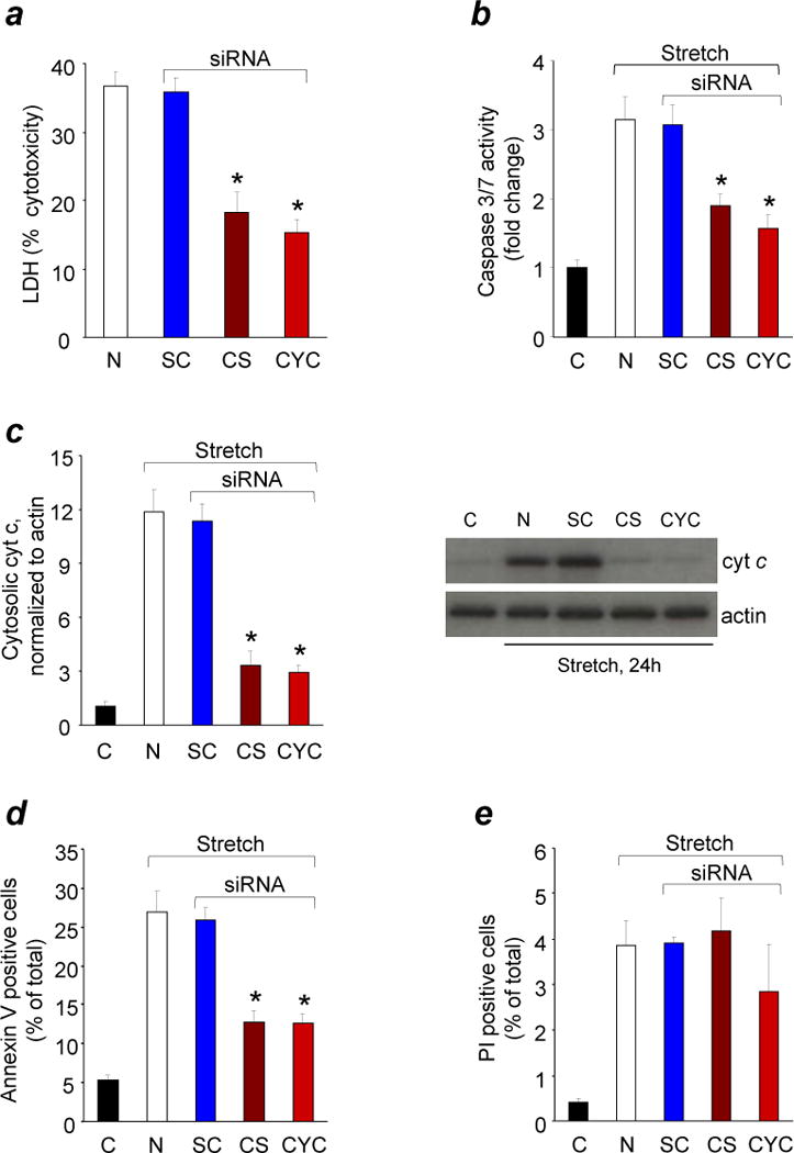
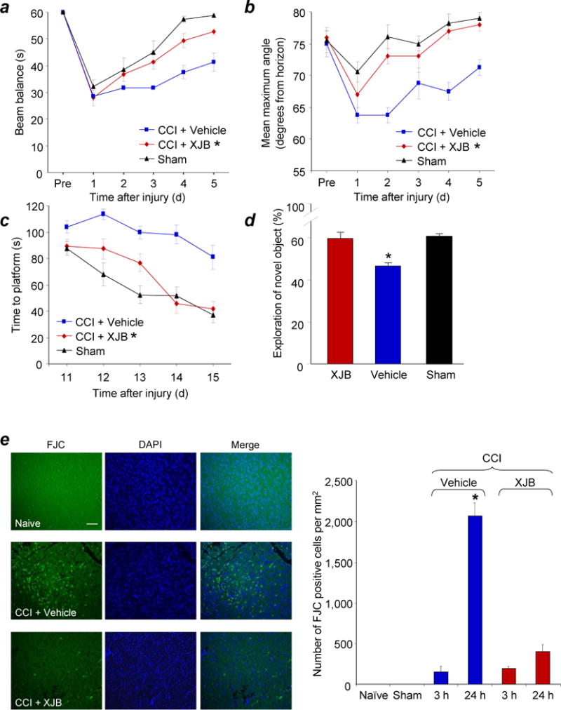
The brain contains a highly diversified complement of molecular species of a mitochondria-specific phospholipid, cardiolipin, which, because of its polyunsaturation, can readily undergo oxygenation. Using global lipidomics analysis in experimental traumatic brain injury (TBI), we found that TBI was accompanied by oxidative consumption of polyunsaturated cardiolipin and the accumulation of more than 150 new oxygenated molecular species of cardiolipin. RNAi-based manipulations of cardiolipin synthase and cardiolipin levels conferred resistance to mechanical stretch, an in vitro model of traumatic neuronal injury, in primary rat cortical neurons. By applying a brain-permeable mitochondria-targeted electron scavenger, we prevented cardiolipin oxidation in the brain, achieved a substantial reduction in neuronal death both in vitro and in vivo, and markedly reduced behavioral deficits and cortical lesion volume. We conclude that cardiolipin oxygenation generates neuronal death signals and that prevention of it by mitochondria-targeted small molecule inhibitors represents a new target for neuro-drug discovery.


Figures

Figure 1
Figure 1
Assessment of molecular species of CL and its oxidation products by 2D-LCMS after TBI. (a) Typical spectra of CL obtained from brain cortex. Upper panel shows the presence of multiple non-oxidized (8-10 major clusters of mass ions shown in blue) and few oxidized (shown in red, CLox) clusters of CL in naïve rat. Left upper insert: 1st dimension chromatographic separation of phospholipids in lipid extracts of the ipsilateral cortex. CL eluted with the 10–12 min retention time window. Right inserts: 2nd dimension chromatographic separation of non-oxidized and oxidized CL. The latter eluted during the 5-6 min retention time window. Middle panel demonstrates non-oxidized (blue) and the appearance of numerous oxidized (red) CL species after TBI. Lower panel illustrates the effect of XJB-5-131 administration after TBI on the profile of non-oxidized (blue) and oxidized (red) CL species. (b) Evaluation of the number of non-oxidized and oxidized molecular species of CL in the brain.(c) Quantification of CL oxidation by 2D-LCMS. Increased content of CLox at 3 h after CCI and its attenuation by XJB-5-131. *P < 0.05 vs. naïve and XJB-5-131; error bars, standard deviation, n = 4 rats per group.
Figure 2
Figure 2
Response of CL or cyt c deficient neurons to in vitro TBI. Quantification of cytotoxicity (lactate dehydrogenase (LDH) release) relative to Triton exposure (a), caspase 3/7 activity (b), cyt c release from mitochondria into cytosol (c), annexin V (d) and propidium iodide (PI) positivity (e) in rat cortical neurons transfected with Cardiolipin synthase (CS) or Cytochrome c (CYC) or scrambled control (SC) siRNA after mechanical stretch. Rat cortical neurons were transfected 72 h before mechanical stretch and measurements were obtained at 24 h after stretch injury. C: control normal neurons; N: non-transfected neurons.* P < 0.01 vs. N and SC; error bars, standard deviation; n = 4 experiments. Stretch induced PI positivity did not change in CL and cyt c deficient neurons (P > 0.05).
Figure 3
Figure 3
Neuronal cell death in response to non-oxidized and oxidized cardiolipin. (a) Quantification of Annexin V and/or propidium iodide (PI) positivity, (b) cytotoxicity (lactate dehydrogenase (LDH) release) relative to Triton exposure, and (c) caspase 3/7 activity in rat cortical neurons exposed to non-oxidized tetralinoleyl cardiolipin (TLCL) and oxidized TLCL (TLCLox) in the form of liposomes. At three tested concentrations (5, 10, 25uM), TLCLox caused dose-dependent cell death in contrast to non-oxidized TLCL. * P < 0.01 vs. 0 μM and TLCL; error bars, standard deviation; n = 3 experiments.
Figure 4
Figure 4
Analysis of XJB-5-131 distribution in neurons and brain. (a) XJB-5-131 (10 μM) partitions into mitochondria in primary cortical neurons. Recovered nitroxide radicals in whole cells, mitochondria, and cytosol fractions were suspended in phosphate buffered saline in the presence or absence of 1.5 mM ferricyanide (K3Fe(CN)6). Insert: representative EPR spectra of XJB-5-131 in different fractions in the presence of ferricyanide. *P < 0.05 vs. without ferricyanide; error bars, standard deviation; n = 4 experiments. (b) EPR-based analysis of XJB-5-131 in CSF in naïve rats. A typical ascorbate radical signal (top) is detected by EPR in the absence of ferricyanide. After addition of ferricyanide, a typical nitroxide signal of XJB-5-131 is detected (bottom). (c) Imaging of XJB-5-131 in the brain of naïve rats by L-Band EPR spectroscopy. For optimal positioning of the head, micro-CT was utilized (upper panel). Lower panels demonstrate typical EPR images of XJB-5-131 distribution in the brain obtained at 5 min and 25 min after its i.v. injection (50 mg/kg). Arrows indicate two nitroxide radical standards (2.5 and 5 μL of 10 mM 3-carboxy-proxyl solution) placed in proximal portions of capillary tubes. (d) Distribution of XJB-5-131 in rat brain assessed by mass spectrometry imaging (MSI) and corresponding Hematoxylin-and-Eosin (H&E) staining of the frozen section. XJB-5-131 was detected as the lithium adduct of its hydroxylamine form at 966 m/z in positive mode TOF/TOF MSI with DHB matrix. The white scale bar is 2 mm. The pixels are a heat map with red being the highest intensity.
Figure 5
Figure 5
Assessments of neurobehavioral and histological outcome in PND 17 rats treated with XJB-5-131 after TBI. (a) Ability of rats to remain (seconds) on the beam balance apparatus before and after CCI or sham injury. A repeated measures ANOVA revealed a significant group (F2,15 = 14.452, P = 0.0003), day (F5,75 = 47.631, P < 0.0001), and group by day interaction effect (F10,75 = 2.186, P = 0.028). Bonferroni post hoc analyses revealed that the CCI + XJB group performed significantly better than the CCI + vehicle group (*P = 0.001; error bars, standard error; n = 7-10 rats per group). (b) Maximum angle (degrees) for rats to remain on an inclined platform. A repeated measures ANOVA revealed significant group (F2,15 = 13.164, P = 0.0005), day (F5,75 = 18.085, P < 0.0001), and group by day interaction (F10,75 = 2.902, P = 0.004). Bonferroni post hoc analyses revealed that the TBI + XJB group performed significantly better than the TBI + vehicle group (*P < 0.001; error bars, standard error; n = 7-10 rats per group). (c) Latency (seconds) for rats to locate a hidden (submerged) platform on post-TBI days 11-15. A repeated measures ANOVA revealed significant group (F2,15 = 19.753, P < 0.0001), day (F4,120 = 22.126, P < 0.0001), and group by day interaction (F8,120 = 2.437, P = 0.018). Bonferroni post hoc analyses revealed that the TBI + XJB group performed significantly better than the TBI + vehicle group (P < 0.001; error bars, standard error; n = 7-10 rats per group) and did not differ from the sham controls (P = 0.08). (d) NOR task performance 29 days after sham or CCI injury. TBI + XJB rats exhibited a better 24-hour delay NOR task performance score compared to TBI + vehicle rats (P < 0.001, ANOVA, F2,27 = 14.736; error bars, standard error; n = 9-10 rats per group). (e) Assessment of neurodegeneration by Fluoro-Jade C (FJC) staining. Neurodegeneration observed in the pericontusional area at 24 h after CCI was attenuated by XJB-5-131. The white scale bar is 40 μm. *P < 0.05 vs. naïve and sham controls, CCI 3h + Vehicle, and CCI + XJB-5-131; error bars, standard deviation; n = 4 rats per group.
Figure 6
Figure 6
Response of neurons treated with 4-Amino-TEMPO (4-AT) to in vitro TBI. (a) Percent cytotoxicity (lactate dehydrogenase (LDH) release relative to Triton exposure (corrected for background LDH). (b) 3-(4,5- dimethylthiazol-2-yl)-2,5-diphenyltetrazolium bromide (MTT) as a percentage of normal control conditions. Vehicle or 4-AT was added to the medium 10 min before stretch. Mechanical stretch induced approximately 35% neuronal death assessed by MTT and LDH at 24 hours. *P < 0.01 vs. stretch neurons, error bars, standard deviation; n = 3 experiments.
Figure 7
Figure 7
Electron paramagnetic resonance (EPR) spectroscopy assessment of inter-conversions of nitroxides and hydroxylamines. (a) Effect of Ascorbate, Ascorbate oxidase and Cyt c/H2O2 on the EPR signal of XJB 5-131. A: XJB-5-131 alone in 35 μL of PBS plus 35 μL of DMSO. B: A plus 250 μM Ascorbate, 1 min. C: A plus 250 μM Ascorbate, 5 min. D: A plus 250 μM Ascorbate, 5 min plus ascorbate oxidase (1U, 15 min). E: D plus10 μM Cyt c. F: D plus10 μM Cyt c/200 μM H2O2. (b) Effect of ascorbate, ascorbate oxidase, and NADH on the EPR signal of XJB-5-131 in brain homogenates. A: 35 μL of Brain Homogenate (350 μg protein) in PBS plus 7 μM XJB-5-131 plus 35μL of DMSO. B: A plus Ascorbate Oxidase (2U), 30 min. C: B plus NADH (200 μM), 30 min. D: C plus K3Fe(CN)6 (100 μM). (c) Effect of pH on the EPR signal of XJB-5-131-OH. 50 μL of 7 μM XJB-5-131-OH in DMSO were mixed with 50 μL of 50 μM HEPES pH 7.45 (open bars) or with 50 μL of 50 μM HEPES pH 8.25 (closed bars). Error bars, standard deviation.

Comment in

References

    1. Faul M, Xu L, Wald MM, Coronado VG. Traumatic brain injury in the United States: emergency department visits, hospitalizations, and deaths 2002-2006. Atlanta (GA): Centers for Disease Control and Prevention, National Center for Injury Prevention and Control; 2010.
    1. Thurman DJ, Alverson C, Dunn KA, Guerrero J, Sniezek JE. Traumatic brain injury in the United States: A public health perspective. J Head Trauma Rehabil. 1999;14:602–615. - PubMed
    1. RAND. RAND Invisible Wounds of War Study. 2011 Jun 26; www.rand.org/multi/military/veterans.html.
    1. Ghajar J. Traumatic brain injury. Lancet. 2000;356:923–929. - PubMed
    1. Maas AI, Roozenbeek B, Manley GT. Clinical trials in traumatic brain injury: past experience and current developments. Neurotherapeutics. 2010;7:115–126. - PMC - PubMed

Publication types

MeSH terms