. 2012 Nov 15;430(2):141-50.
doi: 10.1016/j.ab.2012.08.005. Epub 2012 Aug 21.

Engineered domain-based assays to identify individual antibodies in oligoclonal combinations targeting the same protein

Affiliations

Engineered domain-based assays to identify individual antibodies in oligoclonal combinations targeting the same protein

Q Meng et al. Anal Biochem. .

Abstract

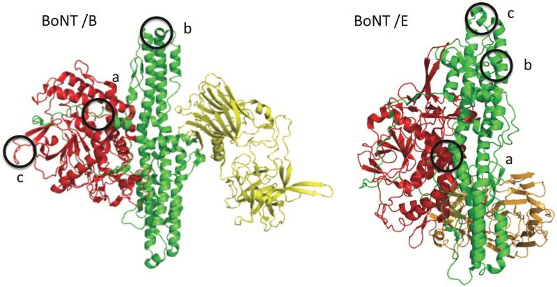
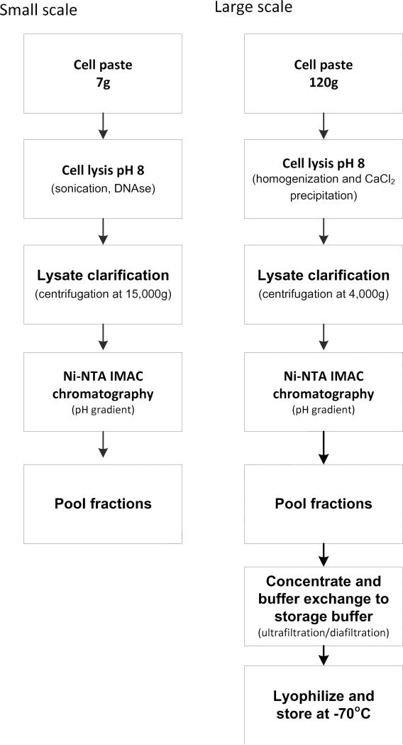
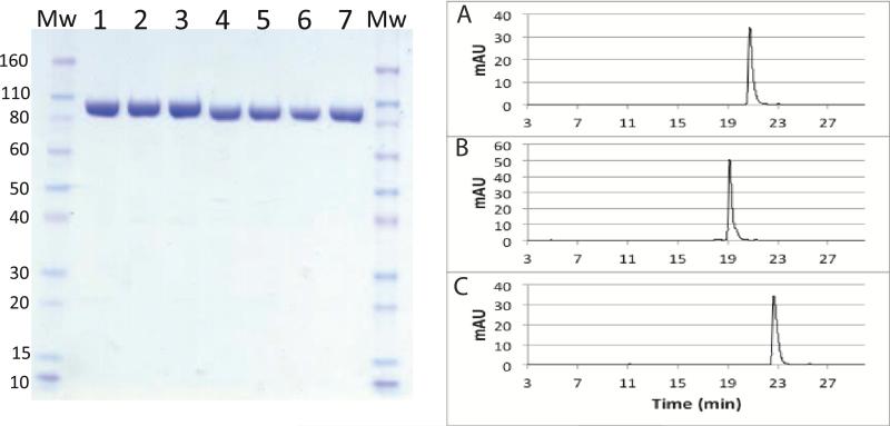
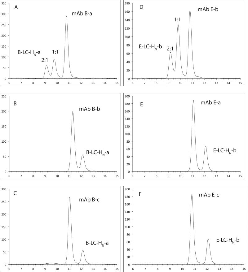
Quantitation of individual monoclonal antibodies (mAbs) within a combined antibody drug product is required for preclinical and clinical drug development. We have developed two antitoxins, XOMA 3B and XOMA 3E, each consisting of three mAbs that neutralize type B and type E botulinum neurotoxin (BoNT/B and BoNT/E) to treat serotype B and E botulism. To develop mAb-specific binding assays for each antitoxin, we mapped the epitopes of the six mAbs. Each mAb bound an epitope on either the BoNT light chain (LC) or translocation domain (H(N)). Epitope mapping data were used to design LC-H(N) domains with orthogonal mutations to make them specific for only one mAb in either XOMA 3B or XOMA 3E. Mutant LC-H(N) domains were cloned, expressed, and purified from Escherichia coli. Each mAb bound only to its specific domain with affinity comparable to the binding to holotoxin. Further engineering of domains allowed construction of enzyme-linked immunosorbent assays (ELISAs) that could characterize the integrity, binding affinity, and identity of each of the six mAbs in XOMA 3B and 3E without interference from the three BoNT/A mAbs in XOMA 3AB. Such antigen engineering is a general method allowing quantitation and characterization of individual mAbs in a mAb cocktail that bind the same protein.


Figures

Figure 1
Figure 1. Epitopes of BoNT/B and BoNT/E mAbs
Left panel: Ribbon cartoon of the X-ray crystal structure of BoNT/B secondary structure (PDB accession code 1S0E) showing the catalytic light chain (LC, red), heavy chain binding domain (HC, yellow) and translocation domain (HN, green). The epitopes of the three mAbs comprising XOMA 3B are circled and labeled a, b, c for each mAb respectively. Right panel: Ribbon cartoon of the X-ray crystal structure of BoNT/E secondary structure (PDB accession code 3FFZ) showing catalytic light chain (LC, red), heavy chain binding domain (HC, yellow) and translocation domain (HN, green). The epitopes of the three mAbs comprising XOMA 3E are circled and labeled a, b, c for each mAb respectively.
Fig.2
Fig.2. Scalable domain purification methods
Small scale (left) and large-scale (right) purification schemes were developed to purify the six BoNT/B and BoNT/E LC-HN domains.
Figure 3
Figure 3. Analysis of the purity of BoNT/B and BoNT/E domains by SDS-PAGE and RP-HPLC
Left panel; A Coomassie-stained SDS-PAGE gel was run to characterize the purity of domains. Molecular weight (MW) markers are at each side of the gel. Lane 1: B-LC-HN-a, lane 2: B-LC-HN-b, lane 3: B-LC-HN-c, lane 4: E-LC-HN-a, lane 5: E-LC-HN-b, lane 6: E-LC-HN-c, lane 7: A-LC-HN-mut. Right panel: Reverse phase high performance liquid chromatography (RP-HPLC) was used to assess domain purity and presence of aggregation. Shown here are three E-LC-HN domain examples: (A) E-LC-HN-a; (B) E-LC-HN-b; (C) E-LC-HN-c.
Figure 4
Figure 4. Analysis of the purity and immunoreactivity of BoNT/B and E domain by size exclusion high performance liquid chromatography (SE-HPLC)
(A-C). When each domain is mixed with its corresponding mAb, the domain peak disappears, indicating that the domain is near 100% immunoreactive. Both 1:1 complex and 2:1 complexed peaks are present. When the domain is mixed with other non-binding mAbs, no complex formation is observed, indicating binding specificity. Shown here are results for two domains: B-LC-HN-a and E-LC-HN-b. (A) B-LC-HN-a with mAb B-a; (B) B-LC-HN-a with mAb B-b; (C) B-LC-HN-a with mAb B-c; (D) E-LC-HN-b with mAb E-b; (E) E-LC-HN-b with mAb E-a; and (F) E-LC-HN-b with mAb E-c.
Figure 5
Figure 5. Direct-coating ELISAs
(A-C), Binding of mAbs B-a, B-b and B-c to plates coated with mAb specific domains B-LC-HN-a (A),B-LC-HN-b (B) or B-LC-HN-c (C). (D-F), Binding of mAbs E-a, E-b, and E-c to plates coated with mAb specific domains E-LC-HN-a (D), E-LC-HN-b (E), or E-LC-HN-c (F). Each mAb bound only to its respective domain, with no binding observed to the other domains.
Figure 6
Figure 6. Specific detection of mAbs A-c and B-b using an indirect capture ELISA
Top panels (A and B): Cartoon of the indirect capture ELISA. Incubation: A fixed amount of mAb specific domain and increasing amounts of mAbs are mixed in individual wells and incubated together until binding reaches equilibrium. Amount of mAb bound is a function of the mAb solution KD. Capture: Each reaction mixture is then transferred to a new well on the capture plate which has been coated with the domain specific mAb for capturing of the domain. Domains bound by a mAb binding the same epitope are not captured and are washed away. Detection: Captured domains are detected with anti-SV5 antibody binding the C-terminal SV5 epitope tag followed by anti-mouse Ab-HRP (A) A-LC-HN mut domain binds mAb A-c with high affinity and mAb E-c with low affinity in solution at a non-overlapping epitope. At the mAb concentrations studied, the affinity of mAb E-c for the A-LC-HN domain is too low for binding to occur during the incubation stage. On the capture plate coated with mAb A-c, only free A-LC-HN mut not bound by mAb E-c is captured, allowing quantitation of the mAb E-c concentration. (B). The B-LC-HN-b domain binds both mAb B-b and mAb E-c with overlapping epitopes, and with a 500-fold higher affinity for mAb B-b than to mAb E-c. At the mAb concentrations studied, the affinity of mAb E-c for the B-LC-HN-b domain is too low for binding to occur during the incubation stage. On the capture plate coated with mAb B-b, the free B-LC-HN-b is captured, while B-LC-HN-b bound by mAb B-b in solution is washed away, allowing quantitation of the mAb E-c concentration. HRP = horse radish peroxidase. Bottom panel (C). Binding specificity of direct vs. indirect capture ELISAs. C1. Direct ELISA with a plate coated with domain A-LC-HN mut shows binding of mAb E-c at higher mAb concentrations than required for mAb A-c. C2 Indirect capture ELISA showed specificity of domain A-LC-HN-mut for only mAb A-c. mAb A-c was coated on the plate as capture antibody, A-LC-HN-mut was incubated with either mAb A-c, or a mixture of eight mAbs (mAbA-a + mAbA-b + XOMA 3B + XOMA 3E) or a mixture of nine mAbs (XOMA 3AB + XOMA 3B + XOMA 3E). C3. Direct ELISA with a plate coated with domain B-LC-HN-b cannot distinguish between mAb B-b and mAb E-c. C4. Indirect capture ELISAs showing specificity of domains B-LC-HN-b for only mAb B-b. mAb B-b was coated on the plate as capture antibody, B-LC-HN-b was incubated with mAb B-b, a mixture of eight mAbs (XOMA 3 AB and mAb B-a, -b and XOMA 3E) or a mixture of nine mAbs (XOMA 3AB + XOMA 3B + XOMA 3E).

References

    1. Nelson AL, Dhimolea E, Reichert JM. Development trends for human monoclonal antibody therapeutics. Nat Rev Drug Discov. 2010;9:767–774. - PubMed
    1. Logtenberg T. Antibody cocktails: next-generation biopharmaceuticals with improved potency. Trends Biotechnol. 2007;25:390–394. - PubMed
    1. Bakker AB, Python C, Kissling CJ, Pandya P, Marissen WE, Brink MF, Lagerwerf F, Worst S, van Corven E, Kostense S, Hartmann K, Weverling GJ, Uytdehaag F, Herzog C, Briggs DJ, Rupprecht CE, Grimaldi R, Goudsmit J. First administration to humans of a monoclonal antibody cocktail against rabies virus: safety, tolerability, and neutralizing activity. Vaccine. 2008;26:5922–5927. - PubMed
    1. Prabakaran M, Prabhu N, He F, Hongliang Q, Ho HT, Qiang J, Meng T, Goutama M, Kwang J. Combination therapy using chimeric monoclonal antibodies protects mice from lethal H5N1 infection and prevents formation of escape mutants. PLoS One. 2009;4:e5672. - PMC - PubMed
    1. Chen Z, Moayeri M, Crown D, Emerson S, Gorshkova I, Schuck P, Leppla SH, Purcell RH. Novel chimpanzee/human monoclonal antibodies that neutralize anthrax lethal factor, and evidence for possible synergy with anti-protective antigen antibody. Infect Immun. 2009;77:3902–3908. - PMC - PubMed

Publication types

MeSH terms