. 2012 Nov;41(5):1577-86.
doi: 10.3892/ijo.2012.1594. Epub 2012 Aug 21.

STAT3 signal transduction through interleukin-22 in oral squamous cell carcinoma

Affiliations

STAT3 signal transduction through interleukin-22 in oral squamous cell carcinoma

Lutfun Naher et al. Int J Oncol. 2012 Nov.

Abstract

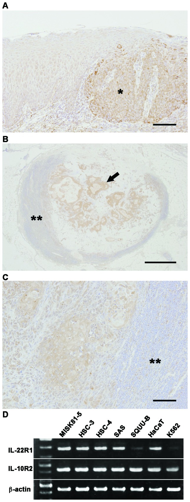
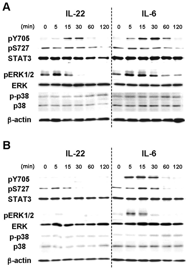
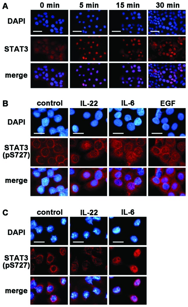
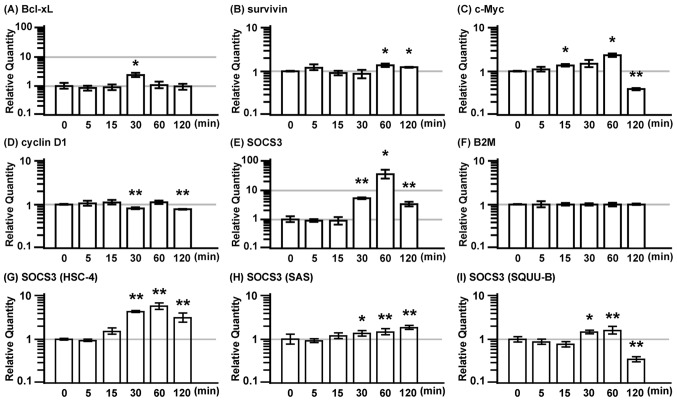
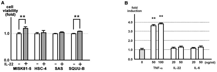
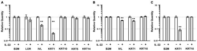
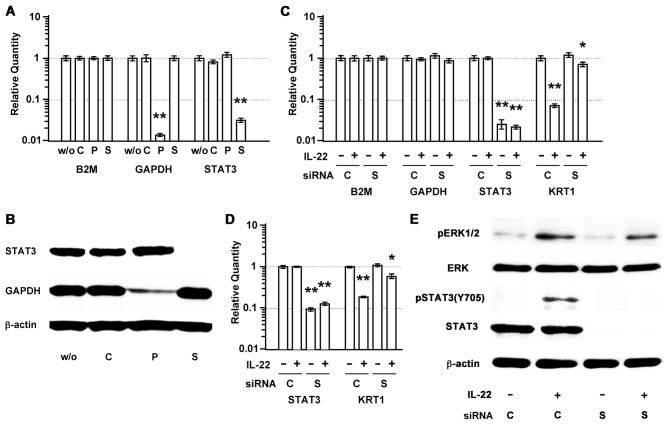
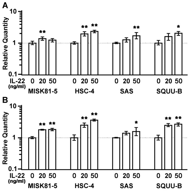
Interleukin (IL)-22 is a member of the IL-10 family. Its main targets are epithelial cells, not immune cells. We examined IL-22 signal transduction in oral squamous cell carcinoma (OSCC) cells. Immunohistochemical staining revealed that IL-22R was expressed more highly in OSCC compared to normal regions. An IL-22R signal was also observed in metastatic OSCC cells in the lymph node. RT-PCR showed that the human OSCC cell lines MISK81-5, HSC-3, HSC-4, SAS and SQUU-B expressed IL-22 receptor chains. Immunoblotting showed that IL-22 induced a transient tyrosine phosphorylation of STAT3 (pY705-STAT3) in MISK81-5 cells. The change in the serine phosphorylation of STAT3 was subtle during the examination periods. Simultaneously, pY705-STAT3 activation in HSC-3 cells was undetectable after IL-22 stimulation. Immunocytochemistry demonstrated that IL-22 induced the translocation of phosphorylated STAT3 into the nucleus of MISK81-5 cells. IL-22 temporarily upregulated the expression of anti-apoptotic and mitogenic genes such as Bcl-x, survivin and c-Myc, as well as SOCS3. IL-22 transiently activated ERK1/2 and induced a delayed phosphorylation of p38 MAP kinase, but negligibly involved the activation of NF-κB in MISK81-5 cells. MISK81-5 and SQUU-B cells treated with IL-22 showed mild cellular proliferation. MISK81-5, HSC-4 and SAS cells treated with IL-22 downregulated the keratinocyte differentiation-related genes compared with unstimulated cells. Conversely, STAT3 suppression by STAT3 siRNA strongly disrupted the downregulation of these genes by IL-22, but it did not significantly affect the activation of ERK1/2 by IL-22. The OSCC cells used in this study upregulated the expression of SERPINB3/4 (SCCA1/2), well-known SCC markers, following treatment with IL-22. These results indicate that IL-22 differentially activates the STAT3 signaling system depending on the type of OSCC. IL-22 may therefore play a role in tumor growth, cell differentiation and progression through STAT3-dependent and -independent pathways.


Figures

Figure 1.
Figure 1.
IL-22R is expressed in the OSCC of the extirpated samples and OSCC cell lines. (A) IL-22R immunostaining was observed in four of the seven extirpated samples, including some normal regions. The immunoreactive intensity for IL-22 was increased in the OSCC region (right side; *) compared with that in the normal oral epithelium (left side). Scale bar, 100 μm. (B) Metastatic carcinoma cells in the cervical lymph node (**) showed strongly positive signals for IL-22R (arrow). Scale bar, 1 mm. (C) IL-22R positive signals in metastatic carcinoma cells in the cervical lymph node (**) in another case. Scale bar, 100 μm. (D) IL-10R2 and IL-22R1 mRNA expressions were examined by semiquantitative RT-PCR. β-actin was used as an internal control.
Figure 2.
Figure 2.
IL-22 induces transient STAT3 phosphorylation and the phosphorylation of several members of the MAPK pathways in MISK81-5, oral squamous cell carcinoma cells. (A) MISK81-5 and (B) HSC-3 cells were incubated with IL-22 (20 ng/ml) or IL-6 (20 ng/ml) for varying times up to 120 min. (A) IL-22 and IL-6 induced pY705-STAT3 in MISK81-5 cells within 15 and 5 min, respectively. IL-22 induced transient tyrosine phosphorylation of STAT3 in MISK81-5 cells with similar kinetics to IL-6. IL-22 and IL-6 induced subtle changes in pS727-STAT3 within the tested time periods. IL-22 induced a transient activation of ERK1/2 in MISK81-5 cells and also induced a delayed phosphorylation of p38 MAP kinase, similar to IL-6. (B) In HSC-3 cells, pY705-STAT3 was undetectable after IL-22 stimulation, although transient pY705-STAT3 expression was induced after IL-6 stimulation. IL-22 and IL-6 induced the transient serine phosphorylation of STAT3. HSC-3 cells stimulated with IL-6 showed activation of ERK1/2 and p38 MAP kinases similar to that in MISK81-5 cells. However, the activation in HSC-3 cells was undetectable after IL-22 treatment. All findings represent the results of three independent experiments.
Figure 3.
Figure 3.
IL-22 translocates pSTAT3 into the nucleus in MISK81-5 cells. (A) MISK81-5 cells were either untreated as a control (left) or treated with IL-6. IL-6 led to the rapid translocation of most of the STAT3 into the nucleus of MISK81-5 cells within 5 min. After 30 min, STAT3 was noted in the cytoplasm again. (B) MISK81-5 or (C) HSC-3 cells were either untreated as a control (left) or treated with IL-22 or IL-6 for 30 min. (B) The translocation of pSTAT3 into the nucleus was noted in MISK81-5 cells treated with IL-22 as well as with IL-6 or EGF. (C) In HSC-3 cells, the translocation of pSTAT3 into the nucleus was not observed after IL-22 stimulation, but trans-location was induced by IL-6. Alexa Fluor® IgG was used as the secondary antibody (red); nuclei were counter-stained with DAPI (blue); EGF was used as control for stimulating the activation of STAT3. (A) Scale bar, 30 μm; (B and C) scale bar, 10 μm.
Figure 4.
Figure 4.
IL-22 transiently induces the expression of anti-apoptotic and mitogenic genes in MISK81-5 cells. MISK81-5 cells were treated with IL-22 (20 ng/ml) for various times as indicated. (A) Bcl-xL, (B) survivin, (C) c-Myc, (D) cyclin D1 and (E) SOCS3 were targeted as STAT3-downstream genes. Bcl-xL, c-Myc and SOCS3 exhibited a peak expression at 30, 60 and 60 min after IL-22 stimulation, respectively. However, c-Myc gene expression was significantly decreased at 120 min. IL-22 significantly increased the gene expression of survivin at 60 and 120 min and decreased cyclin D1 at 30 and 120 min. (F) B2M was used as a reference gene. The SOCS gene expression in (G) HSC-4, (H) SAS and (I) SQUU-B cells treated with IL-22 (20 ng/ml) for various times is shown as indicated. Significant differences between the stimulated and unstimulated samples are indicated with single or double asterisks (*p<0.05; **p<0.01).
Figure 5.
Figure 5.
IL-22 affects MISK81-5 cell proliferation in vitro, but is negligibly associated with upregulation of the cellular NF-κB activation. (A) MISK81-5, HSC-4, SAS and SQUU-B cells were incubated with or without IL-22 (20 ng/ml) for 48 h. The viability of MISK81-5 and SQUU-B cells treated with IL-22 was increased by 1.3- and 1.1-fold in comparison to the control cells, respectively. A significant difference was noted between the stimulated and control samples (**p<0.01). (B) Stably transfected cells, MISK-pGL4-NF-κB cells were stimulated with TNF-α (50 or 100 ng/ml), IL-6 (20 or 50 ng/ml) or IL-22 (20 or 50 ng/ml) or with control medium. A luciferase assay was performed after 24 h. Each ratio is indicated as the relative expression compared to the unstimulated control samples, and indicates the mean ± SD (bars) of three values from one representative experiment. Significant differences in the transcriptional activity are indicated with double asterisks (**p<0.01).
Figure 6.
Figure 6.
IL-22 treatment reduces the expression of keratinocyte differentiation-related genes. (A) The expression of loricrin (LOR), involucrin (IVL), keratin 1 (KRT1), keratin 10 (KRT10), keratin 5 (KRT5) and keratin 14 (KRT14) was compared between MISK81-5 cells stimulated with IL-22 for 24 h and unstimulated cells. (B) The expression of IVL, KRT1 and KRT10 was compared between HSC-4 cells stimulated with IL-22 for 24 h and unstimulated cells. (C) The expression of KRT1 was significantly decreased in the IL-22-treated SAS cells compared to that in unstimulated cells. Significant differences in the gene expression are indicated by double asterisks (**p<0.01).
Figure 7.
Figure 7.
STAT3 siRNA inhibits IL-22-induced reduction of KRT1 expression, but it has little impact on pERK. (A) siRNA selectively reduced the gene expression in the MISK81-5 cells at 30 h after siRNA transfection. Significant differences in the gene expression are indicated by double asterisks (**p<0.01). B2M was used as a reference gene. (B) An immunoblot analysis also revealed that GAPDH and STAT3 siRNAs cause a depletion of the GAPDH and STAT3 protein levels in the MISK81-5 cells, respectively. (C) At 30 h before IL-22 stimulation, the MISK81-5 cells were transfected with siRNA. The expression of B2M, GAPDH, STAT3 and KRT1 was compared between MISK81-5 cells after IL-22 stimulation for 24 h and unstimulated cells (*p<0.05; **p<0.01). (D) The downregulation of KRT1 expression by IL-22 was inhibited in the SAS cells transfected with a siRNA for STAT3 and in unstimulated cells. Significant differences in the gene expression are indicated by single or double asterisks (*p<0.05; **p<0.01). (E) At 30 h after siRNA transfection, the MISK81-5 cells were treated with IL-22 for 10 min. Total cell lysates (10 μg/sample) were analyzed by immunoblotting with an antibody against pY705-STAT3. The membrane was repeatedly reprobed and immunoblotted with an anti-pERK1/2, anti-total STAT3, or an anti-ERK antibody and then with an anti-β-actin antibody. w/o, sample without siRNA treatment; C, sample treated with negative control siRNA; P, sample treated with GAPDH siRNA as a positive control; S, sample treated with STAT3 siRNA.
Figure 8.
Figure 8.
IL-22 treatment induces the upregulated expression of SERPINB3/4 genes in OSCC cells. The expression of the (A) SERPINB3 and (B) SERPINB4 genes was increased in the MISK81-5, HSC-4, SAS and SQUU-B cells stimulated with IL-22 (20 or 50 ng/ml) compared with those cultured in the control medium. Significant differences in the gene expression are indicated by single or double asterisks (*p<0.05; **p<0.01).

Similar articles

Cited by

References

    1. Chidzonga MM, Mahomva L. Squamous cell carcinoma of the oral cavity, maxillary antrum and lip in a Zimbabwean population: a descriptive epidemiological study. Oral Oncol. 2006;42:184–189. - PubMed
    1. Chidzonga MM. Oral malignant neoplasia: a survey of 428 cases in two Zimbabwean hospitals. Oral Oncol. 2006;42:177–183. - PubMed
    1. Dimery IW, Hong WK. Overview of combined modality therapies for head and neck cancer. J Natl Cancer Inst. 1993;85:95–111. - PubMed
    1. Schantz SP, Yu GP. Head and neck cancer incidence trends in young Americans, 1973–1997, with a special analysis for tongue cancer. Arch Otolaryngol Head Neck Surg. 2002;128:268–274. - PubMed
    1. Llewellyn CD, Linklater K, Bell J, Johnson NW, Warnakulasuriya KA. Squamous cell carcinoma of the oral cavity in patients aged 45 years and under: a descriptive analysis of 116 cases diagnosed in the South East of England from 1990 to 1997. Oral Oncol. 2003;39:106–114. - PubMed

Publication types

MeSH terms