Comparative Study
. 2013 Feb;26(1):115-26.
doi: 10.1007/s10334-012-0328-5. Epub 2012 Aug 26.

Quantitative accuracy of attenuation correction in the Philips Ingenuity TF whole-body PET/MR system: a direct comparison with transmission-based attenuation correction

Affiliations
Comparative Study

Quantitative accuracy of attenuation correction in the Philips Ingenuity TF whole-body PET/MR system: a direct comparison with transmission-based attenuation correction

Georg Schramm et al. MAGMA. 2013 Feb.

Erratum in

  • MAGMA. 2015 Feb;28(1):101

Abstract

Objective: Evaluation of the quantitative accuracy of MR-based attenuation correction (MRAC) in the Philips Ingenuity TF whole-body PET/MR.

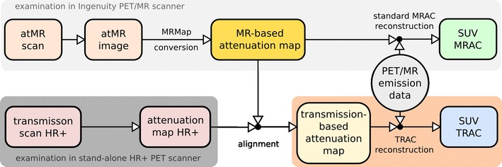
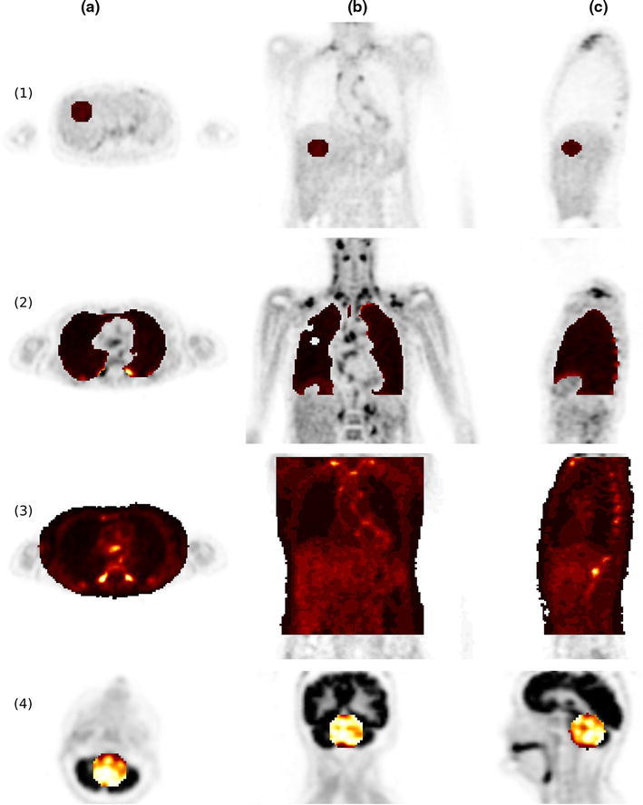
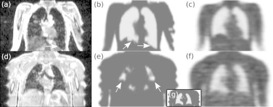
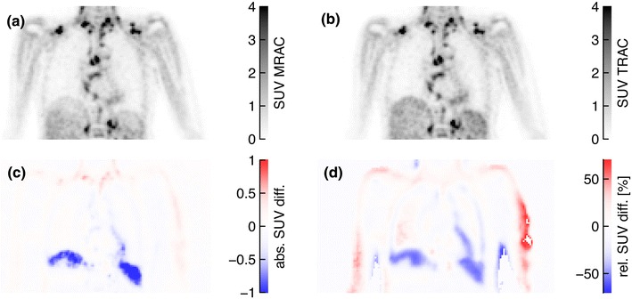
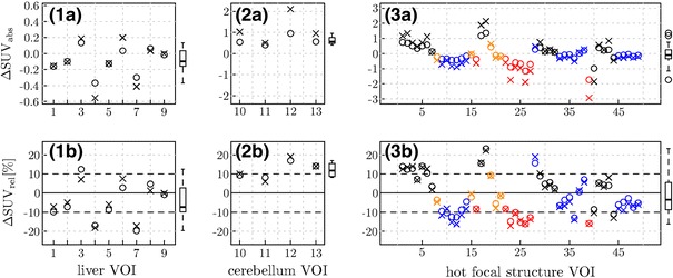
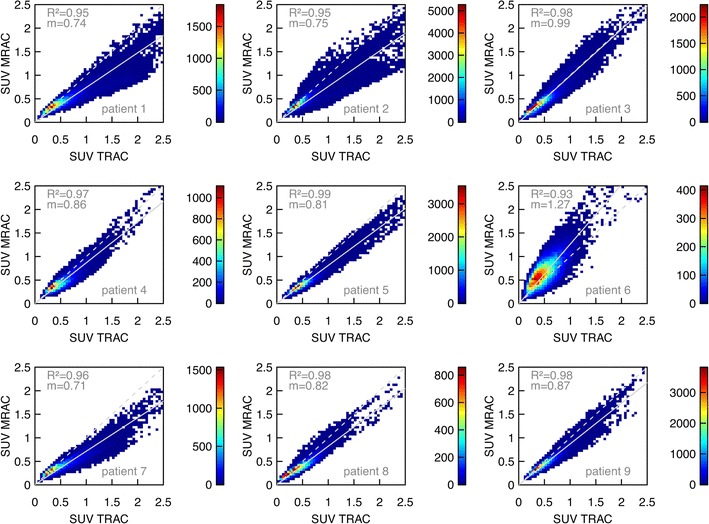
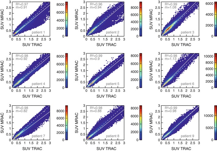
Materials and methods: In 13 patients, PET emission data from the PET/MR were reconstructed using two different methods for attenuation correction. In the first reconstruction, the vendor-provided standard MRAC was used. In the second reconstruction, a coregistered transmission-based attenuation map from a second immediately preceding investigation with a stand-alone Siemens ECAT EXACT HR(+) PET scanner was used (TRAC). The two attenuation maps were compared regarding occurrence of segmentation artifacts in the MRAC procedure. Standard uptake values (SUVs) of multiple VOIs (liver, cerebellum, hot focal structures at various locations in the trunk) were compared between both reconstructed data sets. Furthermore, a voxel-wise intensity correlation analysis of both data sets in the lung and trunk was performed.

Results: VOI averaged SUV differences between MRAC and TRAC were as follows (relative differences, mean ± standard deviation): (+12 ± 6) % cerebellum, (-4 ± 9) % liver, (-2 ± 11) % hot focal structures. The fitted slopes of the voxel-wise correlations in the lung and trunk were 0.87 ± 0.17 and 0.95 ± 0.10 with averaged adjusted R (2) values of 0.96 and 0.98, respectively. These figures include two instances with partially erroneous lung segmentation due to artifacts in the underlying MR images.

Conclusion: The MR-based attenuation correction implemented on the Philips Ingenuity PET/MR provides reasonable quantitative accuracy. On average, deviations from TRAC-based results are small (on the order of 10% or below) across the trunk, but due to interindividual variability of the segmentation quality, deviations of more than 20% can occur. Future improvement of the segmentation quality would help to increase the quantitation accuracy further and to reduce the inter-subject variability.


Figures

Fig. 1
Fig. 1
Scheme of the two reconstructions of the PET/MR emission data using MR-based attenuation correction and an external transmission-based attenuation correction. Further details are given in the text
Fig. 2
Fig. 2
Representative transaxial (a), coronal (b) and sagittal (c) views of VOI definition in liver (1), lung (2), trunk (3) and cerebellum (4)
Fig. 3
Fig. 3
Two instances of incorrect lung segmentation. Corresponding slices of atMR (left), MRmap (middle), and TRmap (right) are shown for patient 1 (ac) and 6 (df), respectively. The inset (g) displays the much improved lung segmentation in the latter patient after adjusting the relevant parameter of the segmentation algorithm
Fig. 4
Fig. 4
Corresponding coronal PETMRAC (a) and PETTRAC (b) slices of patient 1. The absolute and relative SUV differences according to Eqs. (1) and (2) are shown in (c) and (d), respectively. In order to improve visibility of the relevant differences, some spurious voxels in the left arm are suppressed in image (d) by choosing a suitable upper threshold
Fig. 5
Fig. 5
Same as Fig. 4 for a sagittal slice of patient 11. In order to improve visibility of the relevant differences, some spurious voxels in the chin are suppressed in image (d) by choosing a suitable upper threshold. Subfigures (e) and (f) show the corresponding slices of the MRmap and TRmap, respectively
Fig. 6
Fig. 6
Absolute (top) and relative (bottom) SUV differences between PETMRAC and PETTRAC in the liver (1), the cerebellum (2), and several hot focal structures (3) located in the lung and mediastinum (red), in the thoracic spine (blue), in the liver and kidneys (orange) and in the clavicular region (black), respectively. Crosses indicate SUVmaxcircles SUVmean deviations in the respective VOIs. The horizontal dashed lines show ±10 % deviations. The boxplots in the right margins of the plots represent the resulting distributions of the respective SUVmean deviations
Fig. 7
Fig. 7
Voxel intensity (SUV) correlation between PETMRAC and PETTRAC in the lungs of patients 1–9. The correlations are displayed as 2D histograms of 50 × 50 bins with color-coded frequencies. The dashed line indicates the line of identity, the solid line represents the best fit of Eq. (3) to the data. The adjusted R 2 and slope m of the fitted straight line through the origin are specified in the plots
Fig. 8
Fig. 8
Same as Fig. 7 for all voxels in the trunk of patients 1–9

References

    1. Zaidi H, Ojha N, Morich M, Griesmer J, Hu Z, Maniawski P, Ratib O, Izquierdo-Garcia D, Fayad Za, Shao L. Design and performance evaluation of a whole-body Ingenuity TF PET-MRI system. Phys Med Biol. 2011;56:3091–3106. doi: 10.1088/0031-9155/56/10/013. - DOI - PMC - PubMed
    1. Delso G, Fürst S, Jakoby B, Ladebeck R, Ganter C, Nekolla SG, Schwaiger M, Ziegler SI. Performance measurements of the Siemens mMR integrated whole-body PET/MR scanner. J Nucl Med. 2011;52:1914–1922. doi: 10.2967/jnumed.111.092726. - DOI - PubMed
    1. Antoch G, Bockisch A. Combined PET/MRI: a new dimension in whole-body oncology imaging? Eur J Nucl Med Mol Imaging. 2009;36(Suppl 1):S113–S120. doi: 10.1007/s00259-008-0951-6. - DOI - PubMed
    1. Heiss WD. The potential of PET/MR for brain imaging. Eur J Nucl Med Mol Imaging. 2009;36(Suppl 1):S105–S112. doi: 10.1007/s00259-008-0962-3. - DOI - PubMed
    1. Nekolla SG, Martinez-Moeller A, Saraste A. PET and MRI in cardiac imaging: from validation studies to integrated applications. Eur J Nucl Med Mol Imaging. 2009;36(Suppl 1):S121–S130. doi: 10.1007/s00259-008-0980-1. - DOI - PubMed

Publication types