. 2013 Nov;23(11):2552-67.
doi: 10.1093/cercor/bhs234. Epub 2012 Aug 23.

The doublesex homolog Dmrt5 is required for the development of the caudomedial cerebral cortex in mammals

Affiliations

The doublesex homolog Dmrt5 is required for the development of the caudomedial cerebral cortex in mammals

Amandine Saulnier et al. Cereb Cortex. 2013 Nov.

Abstract

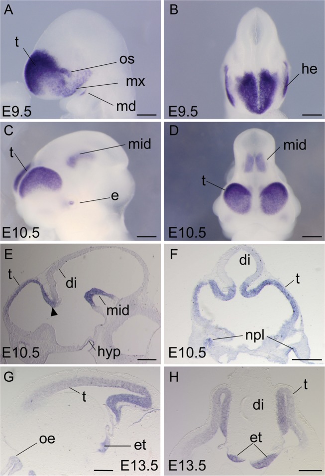
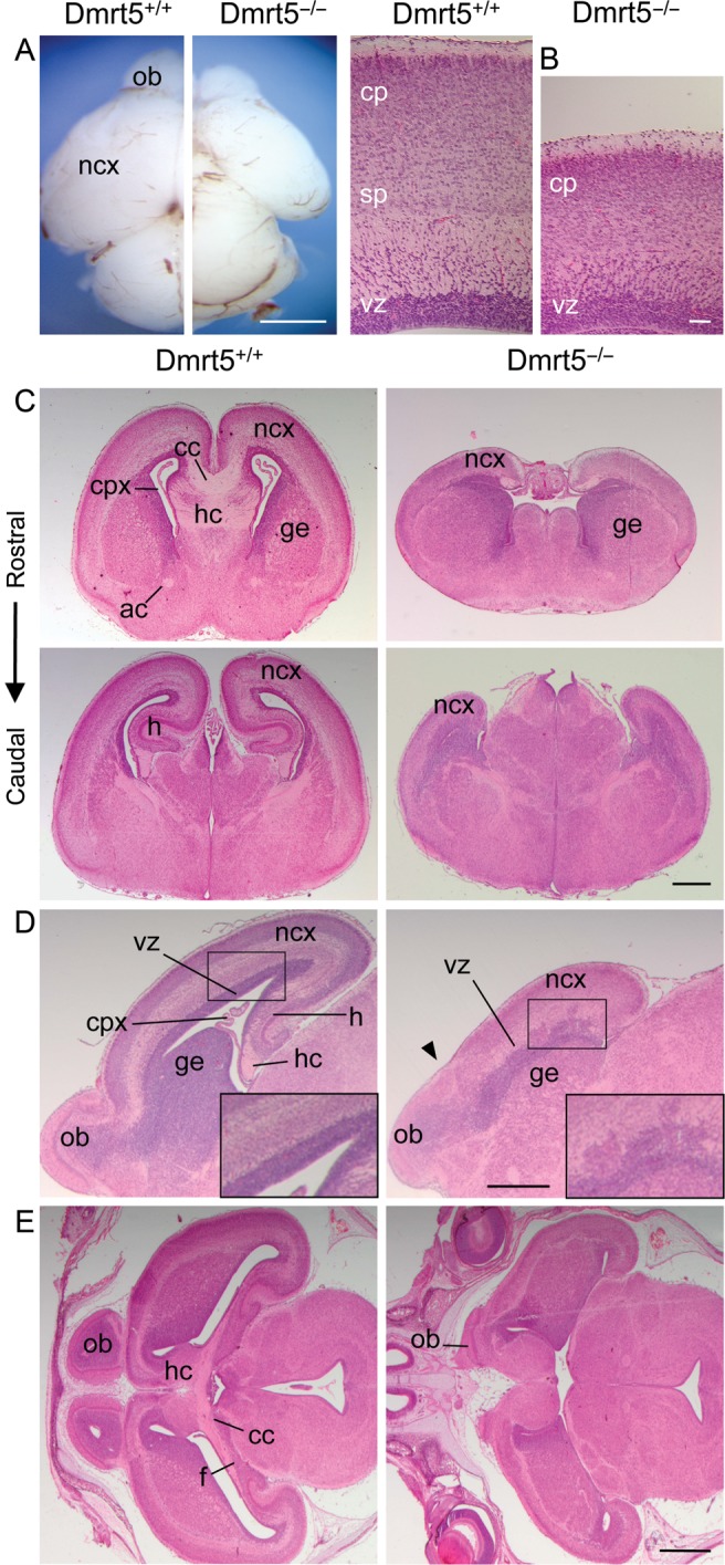
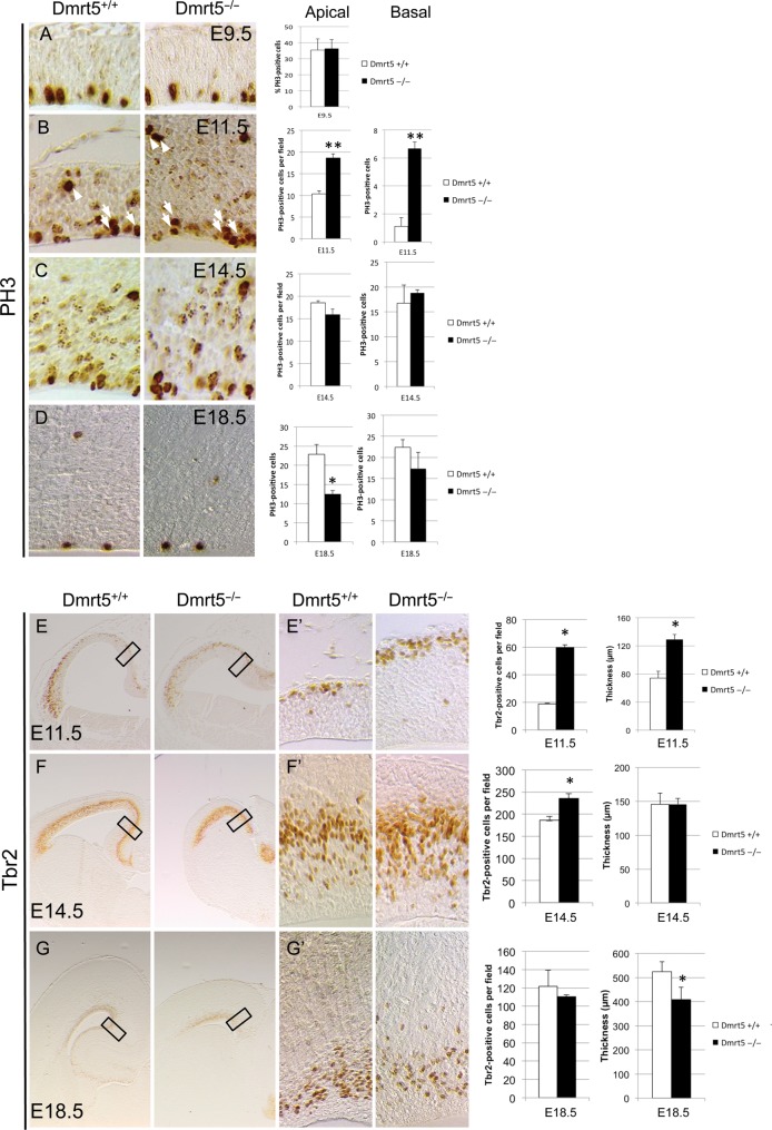
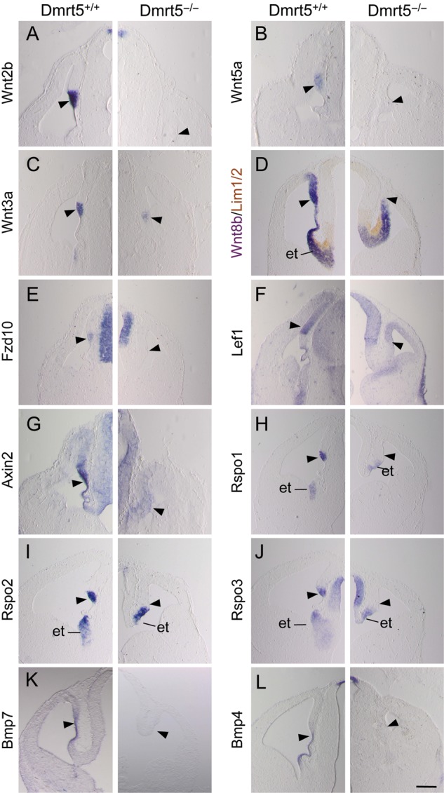
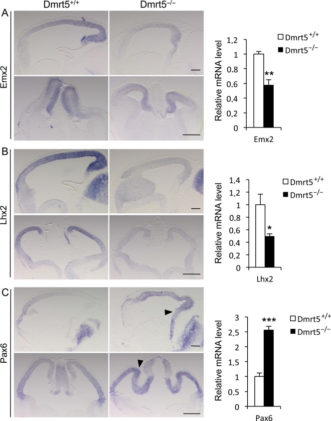
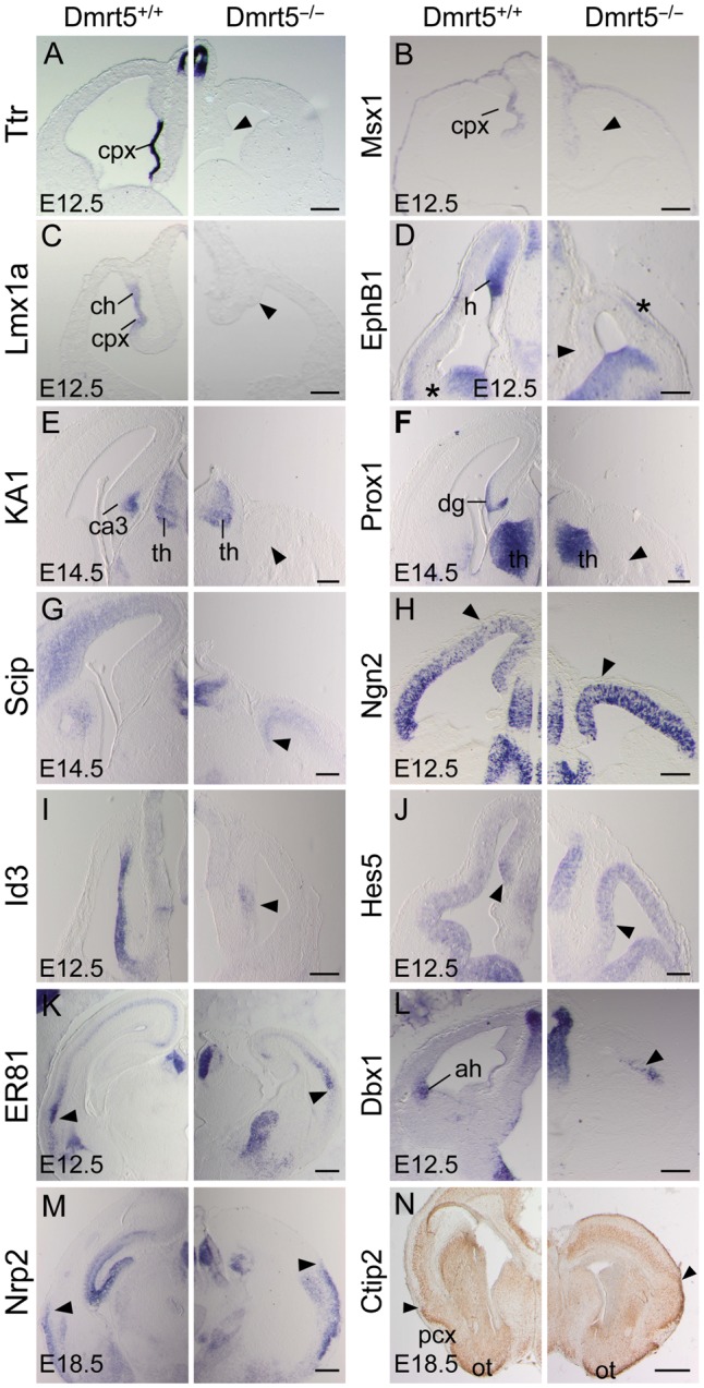
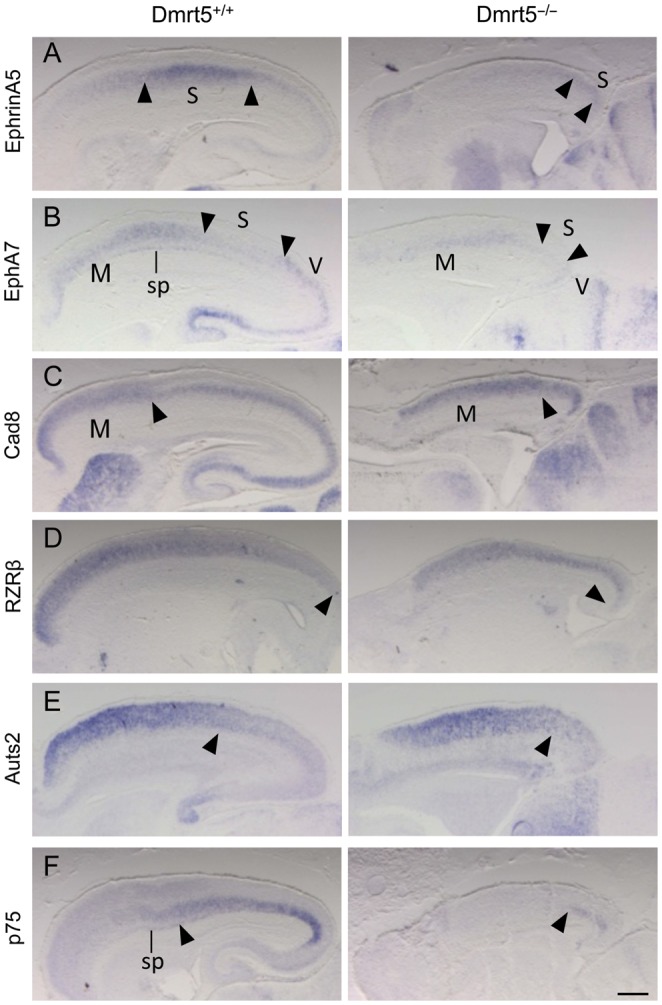
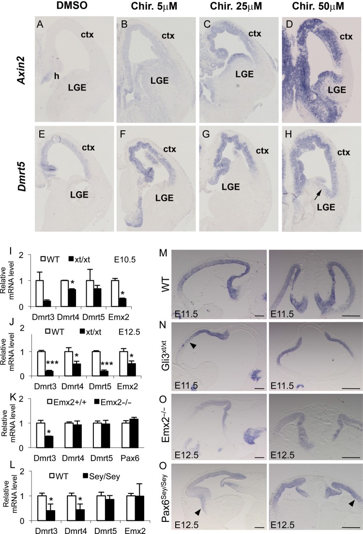
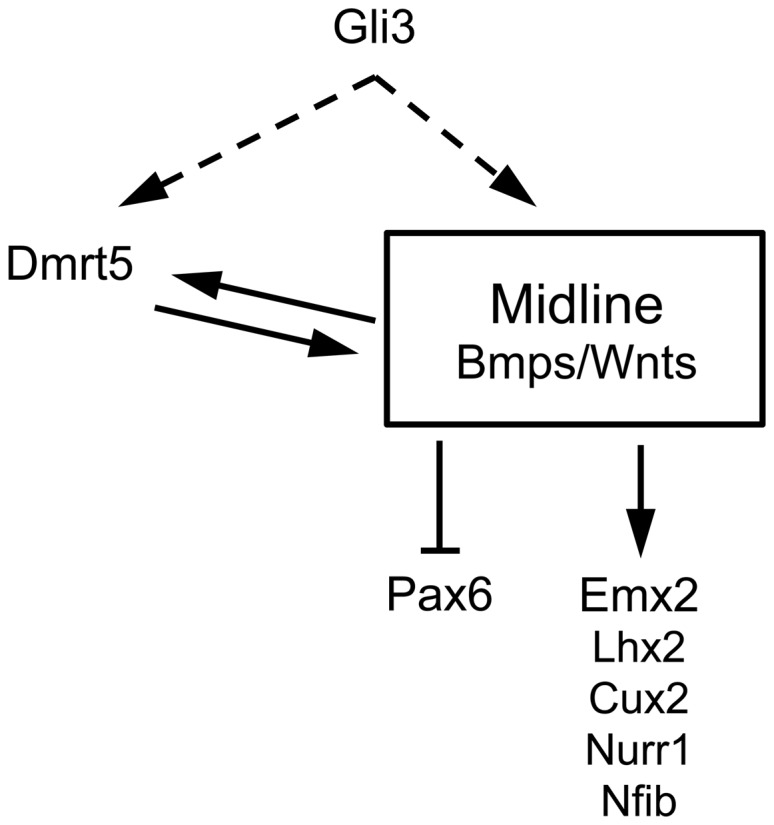
Regional patterning of the cerebral cortex is initiated by morphogens secreted by patterning centers that establish graded expression of transcription factors within cortical progenitors. Here, we show that Dmrt5 is expressed in cortical progenitors in a high-caudomedial to low-rostrolateral gradient. In its absence, the cortex is strongly reduced and exhibits severe abnormalities, including agenesis of the hippocampus and choroid plexus and defects in commissural and thalamocortical tracts. Loss of Dmrt5 results in decreased Wnt and Bmp in one of the major telencephalic patterning centers, the dorsomedial telencephalon, and in a reduction of Cajal-Retzius cells. Expression of the dorsal midline signaling center-dependent transcription factors is downregulated, including Emx2, which promotes caudomedial fates, while the rostral determinant Pax6, which is inhibited by midline signals, is upregulated. Consistently, Dmrt5(-/-) brains exhibit patterning defects with a dramatic reduction of the caudomedial cortex. Dmrt5 is increased upon the activation of Wnt signaling and downregulated in Gli3(xt/xt) mutants. We conclude that Dmrt5 is a novel Wnt-dependent transcription factor required for early cortical development and that it may regulate initial cortical patterning by promoting dorsal midline signaling center formation and thereby helping to establish the graded expression of the other transcription regulators of cortical identity.

Keywords: Emx2; Wnt/Bmp; choroid plexus; cortical hem; telencephalon.


Figures

Figure 1.
Figure 1.
Dmrt5 expression in the mouse developing brain. (AD) Whole-mount ISH of Dmrt5 at E9.5 (A and B) and E10.5 (C and D). (EH) Sagittal (E and G) and coronal (F and H) brain sections. Arrowhead in (E) indicates Dmrt5 expression at the telen-diencephalic boundary. di, diencephalon; e, eye; et, eminentia thalami; he, head ectoderm; hyp, hypophysis; md, mandibular process; mid, midbrain; mx, maxillary process; npl, nasal placode; oe, olfactory epithelium; os, optic stalk; t, telencephalon. Scale bars: 200 μm in A and B; 500 μm in CH.
Figure 2.
Figure 2.
Reduced cerebral cortex with midline defects in Dmrt5−/− mice. (A) Dorsal views of P0 control and Dmrt5−/− brains. (B) High magnification view of sagittal sections of E18.5 control and mutant brains. (CE) Hematoxylin and eosin staining of coronal (C, at the rostral level, upper panels, and the caudal level, lower panels), sagittal (D) and horizontal (E) sections of E18.5 control and mutant brains. Note the abnormal limit of the neocortex with the olfactory bulbs (arrowhead in D). ac, anterior commissure; cc, corpus callosum; cp, cortical plate; cpx, choroid plexus; f, fimbria; ge, ganglionic eminences; h, hippocampus; hc, hippocampal commisure; ncx, neocortex; ob, olfactory bulbs; sp, subplate; vz, ventricular zone. Scale bars: 200 μm in A and B; 500 μm in C and D; 1 mm in E.
Figure 3.
Figure 3.
Altered cell proliferation in the telencephalon of Dmrt5−/− mice. (AD) PH3 IHC on coronal sections of Dmrt5−/− and control embryos at the indicated stages. Quantification of the results is shown on the right. Note the transient slight increased in apical and larger increase in basal PH3 cells at E11.5 and later slight decrease in PH3-positive cells in Dmrt5−/− embryos. (EG) Tbr2 IHC on coronal sections of Dmrt5−/− and control embryos at the indicated stages. High magnifications of the rectangles in (EG) are shown in (E′–G′). Quantification of the results and thickness of the cortical wall is shown on the right. Note the transient increase of the number of Tbr2-positive cells and of the thickness of the cortical wall at E11.5, and the decrease of the thickness at E18.5 in Dmrt5−/− embryos (*P< 0.05 and **P< 0.01). Error bars indicate standard deviation (SD).
Figure 4.
Figure 4.
Reduction of Wnt and Bmp genes and genes encoding Wnt signaling components in the caudomedial cerebral cortical wall of Dmrt5 mutants. (AC and EL) Coronal forebrain sections of mutant and control mice at E12.5 analyzed by ISH for the indicated genes. (D) ISH for Wnt8b combined to immunoperoxidase staining for Lim1/2, a marker of diencephalic cells. Note that all tested genes were undetectectable or strongly reduced in the CH region (arrowheads) of E12.5 mutant mice. et, eminentia thalami. Scale bars: 500 μm.
Figure 5.
Figure 5.
Downregulation of Emx2 and Lhx2 and upregulation of Pax6 in the cortex of E12.5 Dmrt5 mutants. (AC) ISH for the indicated markers on sagittal (upper panels) and coronal (lower panels) sections. Sagittal sections are shown with anterior to the left. The arrowheads in (C) indicate Pax6 upregulation in the medial and posterior part of the cortex of the mutants. Quantitative RT–qPCR analysis of Emx2, Lhx2, and Pax6 in the cortex of WT and mutant E12.5 embryos is shown on the right. Results are normalized to the level of expression in the WT forebrain. Error bars show SD of 3 independent experiments. Scale bars: 200 μm in upper panels; 500 μm in lower panels (*P< 0.05, **P< 0.01, and ***P< 0.001).
Figure 6.
Figure 6.
The Absence of choroid plexus and hippocampal markers and altered pattern of mediolateral regional markers in the dorsal telencephalon of Dmrt5−/− embryos. (AM) Coronal forebrain sections processed by ISH for the indicated markers at the indicated stages. (AJ) While Ttr, Msx1, Lmx1a, EphB1, KA1, Prox1, Scip, Id3, and Hes5 medial markers were strongly reduced in the mutants (arrowheads), Ngn2 expression spreads from the lateral part of the cortex toward the dorsomedial cortex (arrowheads in H). (KM) ER81, Dbx1 and Nrp2 as well as a site of EphB1 expression in the lateral part of the cortex (asterisk in D) appeared all shifted medially in the lateral cortical wall of mutant mice (arrowheads). (N) Olfactory cortex specific Ctip2 expression appears shifted medially while neocortical-specific expression extends less ventrally in the mutant. The limit between the neocortex and piriform cortex is indicated (arrowheads). ah, anti-hem; ch, cortical hem; cpx, choroid plexus; dg, dentate gyrus; h, hippocampal primordium; ot, olfactory tubercles; pcx, piriform cortex; th, thalamus. Scale bars: 500 μm in AM; 1 mm in L.
Figure 7.
Figure 7.
Reduction of caudomedial neocortical areas in Dmrt5 mutants. Sagittal brain sections were analyzed by ISH for the indicated genes. (AC) EphrinA5 domain of high expression and EphA7 domain of low expression that demarcate the somatosensory areas (delimitated by arrowheads) is contracted and detected in the most caudal part of the cortex in Dmrt5−/− embryos. EphA7 and Cad8 high expression that mark the rostrally located motor area (with caudal limits indicated by arrowheads) occupies most of the reduced cortex in mutants. (DF) RZRβ and Auts2 are expanded and p75 constricted caudally in mutants. Arrowheads mark the approximate caudal and rostral limit of their expression, respectively. Note the reduced EphA7 and p75 in the subplate. M, motor; S, somatosensory; sp, subplate; V, visual areas. Scale bars: 500 μm.
Figure 8.
Figure 8.
Dmrt5 expression in the cortex is upregulated by activation of Wnt/β-catenin, downregulated in Gli3xt/xt embryos, and unchanged in Emx2−/− a Pax6sey/sey embryo. (AH) Dmrt5 expression in Chir-treated and control ex vivo cortical explants. Note the activation of Dmrt5 throughout the telencephalic ventricular zone similar to that observed for Axin2 used as a positive control after addition of Chir in a concentration-dependent manner. Note also the Dmrt5 ectopic expression detected at high dose in the ventricular zone of the lateral ganglionic eminence (arrow). (IP) Dmrt5 expression in Gli3xt/xt, Emx2−/−, and Pax6sey/sey embryos. (IL) Quantitative RT–qPCR analysis of Dmrt3, 4, and 5 in the cortex of WT and Gli3xt/xt at E10.5 (I) and E12.5 (J) and in Emx2−/− (K) and Pax6sey/sey (L) at E12.5. Results are normalized to the level of expression in the WT forebrain. Error bars show SD of 3 independent experiments (*P< 0.05 and ***P< 0.001). (MP) Sagittal (left panels) and coronal (right panels) brain sections of WT (M), Gli3xt/xt (N), Emx2−/− (O), and Pax6sey/sey (P) embryos, processed by ISH for Dmrt5 at the indicated stage. Note in (N) the reduction anteriorly and in (P) the slight extension on Dmrt5 anteriorly and laterally (arrowheads). Ctx, cortex; h, hem; LGE, lateral ganglionic eminences. Scale bars in (MP): 200 μm, left panels; 500 μm, rights panels.
Figure 9.
Figure 9.
Summary of the interactions identified in this work between Dmrt5 and the secreted ligands and Wnt/Bmp regulated graded transcription factors expressed during early patterning of the cerebral cortex. Wnt and Gli3 control Dmrt5 expression. Direct or indirect action of Gli3 on Dmrt5, through regulation of the Wnt signaling pathway, is indicated by dashed lines. Dmrt5 in turn is required for Wnt and Bmp expression in the dorsal midline signaling center. In the absence of Dmrt5, this feedback loop is not maintained, which is likely responsible for the downregulation of Wnt target genes. Pax6 is upregulated in Dmrt5 mutants, presumably through its negative regulation by Wnts and Emx2.

Similar articles

Cited by

References

    1. Allendoerfer KL, Shatz CJ. The subplate, a transient neocortical structure: Its role in the development of connections between thalamus and cortex. Annu Rev Neurosci. 1994;17:185–218. - PubMed
    1. Aoto K, Nishimura T, Eto K, Motoyama J. Mouse GLI3 regulates Fgf8 expression and apoptosis in the developing neural tube, face, and limb bud. Dev Biol. 2002;251:320–332. - PubMed
    1. Armentano M, Chou SJ, Tomassy GS, Leingärtner A, O'Leary DD, Studer M. COUP-TF1 regulates the balance of cortical patterning between frontal/motor and sensory areas. Nat Neurosci. 2007;10:1277–1286. - PubMed
    1. Balciuniene J, Bardwell VJ, Zarkower D. Mice mutant in the DM domain gene Dmrt4 are viable and fertile but have polyovular follicles. Mol Cell Biol. 2006;26:8984–8991. - PMC - PubMed
    1. Bedogni F, Hodge RD, Elsen GE, Nelson BR, Daza RA, Beyer RP, Bammler TK, Rubenstein JL, Hevner RF. Tbr1 regulates regional and laminar identity of postmitotic neurons in developing neocortex. Proc Natl Acad Sci USA. 2010;107:13129–13134. - PMC - PubMed

Publication types

Substances