

AS30D model of hepatocellular carcinoma: tumorigenicity and preliminary characterization by imaging, histopathology, and immunohistochemistry
- PMID: 22923329
- PMCID: PMC3904804
- DOI: 10.1007/s00270-012-0466-1
AS30D model of hepatocellular carcinoma: tumorigenicity and preliminary characterization by imaging, histopathology, and immunohistochemistry
Abstract
Purpose: This study was designed to determine the tumorigenicity of the AS30D HCC cell line following orthotopic injection into rat liver and preliminarily characterize the tumor model by both magnetic resonance imaging (MRI) and ultrasound (US) as well as histopathology and immunohistochemistry.
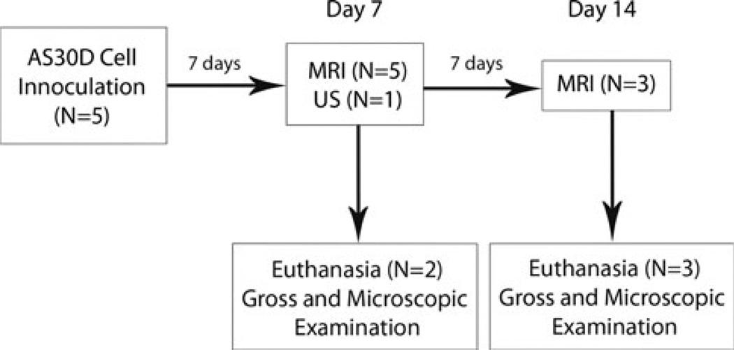
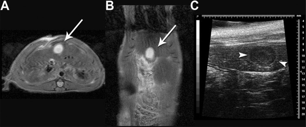
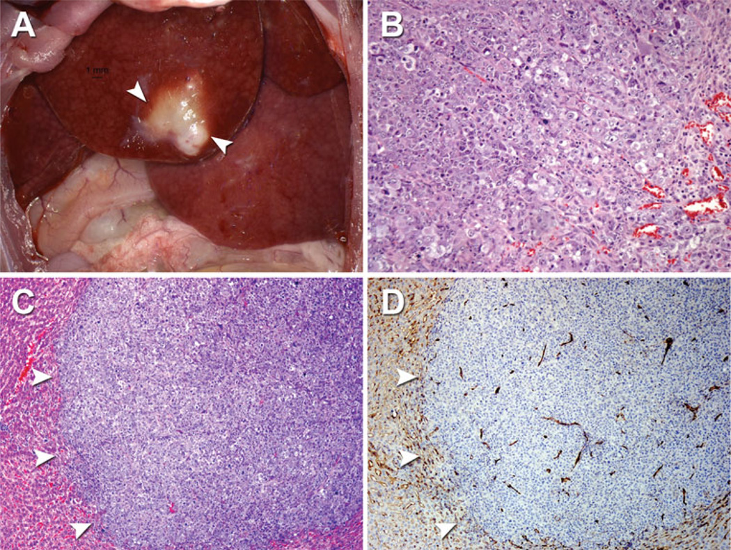
Materials: AS30D cell line in vitro proliferation was assessed by using MTT assay. Female rats (N = 5) underwent injection of the AS30D cell line into one site in the liver. Rats subsequently underwent MR imaging at days 7 and 14 to assess tumor establishment and volume. One rat underwent US of the liver at day 7. Rats were euthanized at day 7 or 14 and livers were subjected to gross, histopathologic (H&E), and immunohistochemical (CD31) analysis to assess for tumor growth and neovascularization.
Results: AS30D cell line demonstrated an in vitro doubling time of 33.2 ± 5.3 h. MR imaging demonstrated hyperintense T2-weighted and hypointense T1-weighted lesions with tumor induction in five of five and three of three sites at days 7 and 14, respectively. The mean (SD) tumor volume was 126.1 ± 36.2 mm(3) at day 7 (N = 5). US of the liver demonstrated a well-circumscribed, hypoechoic mass and comparison of tumor dimensions agreed well with MRI. Analysis of H&E- and CD31-stained sections demonstrated moderate-high grade epithelial tumors with minimal tumor necrosis and evidence of diffuse intratumoral and peritumoral neovascularization by day 7.
Conclusions: AS30D HCC cell line is tumorigenic following orthotopic injection into rat liver and can be used to generate an early vascularizing, slower-growing rat HCC tumor model.
Conflict of interest statement
Figures
References
-
- Aravalli RN, Steer CJ, Sahin MB, Cressman EN. Stem cell origins and animal models of hepatocellular carcinoma. Dig Dis Sci. 2010;55(5):1241–1250. - PubMed
-
- Aravalli RN, Golzarian J, Cressman EN. Animal models of cancer in interventional radiology. Eur Radiol. 2009;19(5):1049–1053. - PubMed
-
- Ju S, McLennan G, Bennett SL, et al. Technical aspects of imaging and transfemoral arterial treatment of N1-S1 tumors in rats: an appropriate model to test the biology and therapeutic response to transarterial treatments of liver cancers. J Vasc Interv Radiol. 2009;20(3):410–414. - PubMed
Publication types
MeSH terms
Grants and funding
LinkOut - more resources
Full Text Sources
Medical
