. 2012 Nov 1;40(20):10345-55.
doi: 10.1093/nar/gks753. Epub 2012 Aug 25.

Dynamic regulation of HIV-1 mRNA populations analyzed by single-molecule enrichment and long-read sequencing

Affiliations

Dynamic regulation of HIV-1 mRNA populations analyzed by single-molecule enrichment and long-read sequencing

Karen E Ocwieja et al. Nucleic Acids Res. .

Abstract

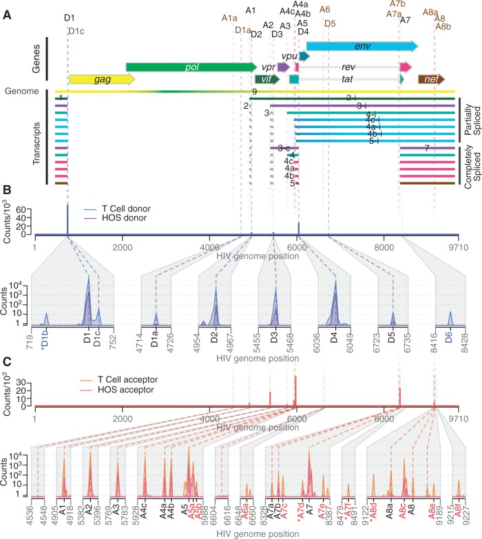
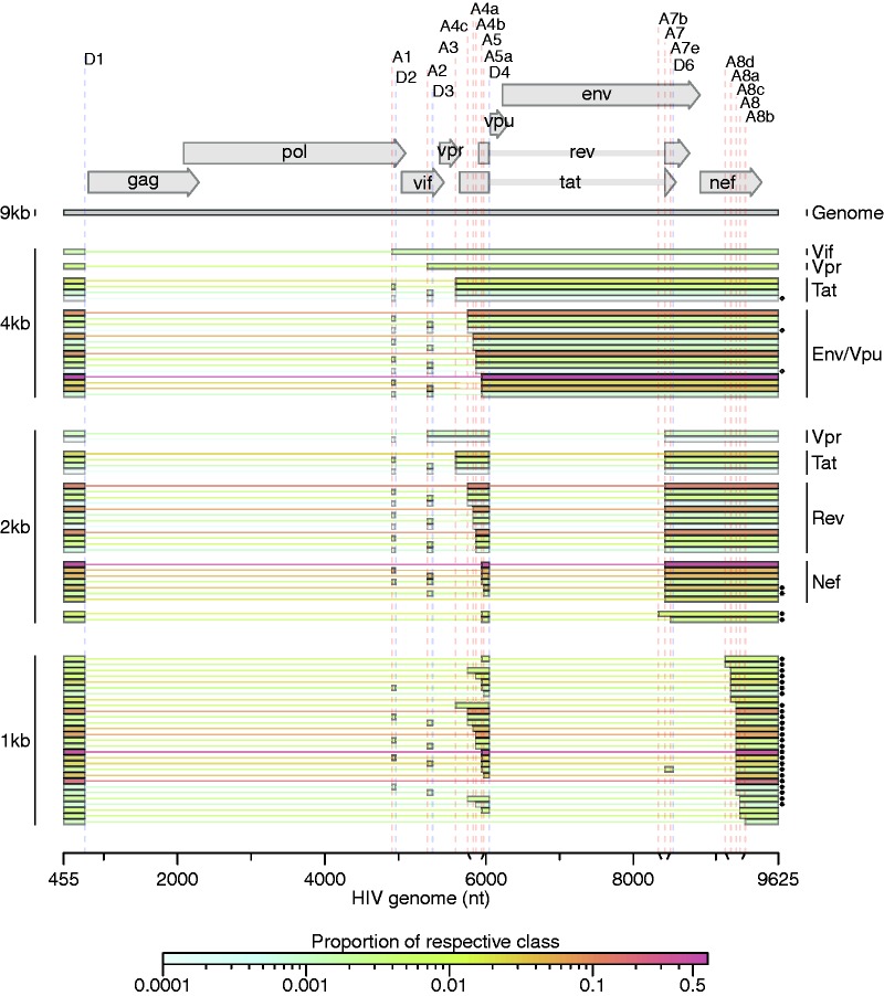
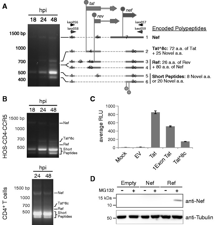
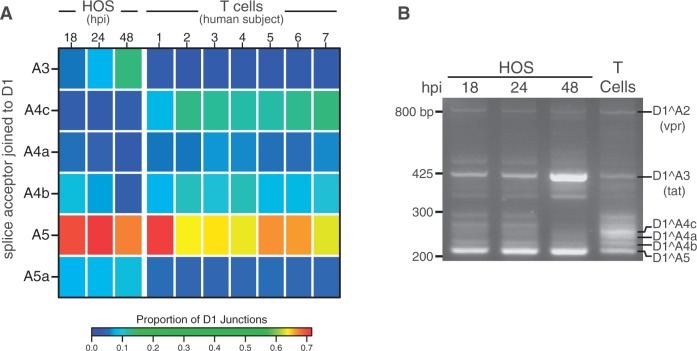
Alternative RNA splicing greatly expands the repertoire of proteins encoded by genomes. Next-generation sequencing (NGS) is attractive for studying alternative splicing because of the efficiency and low cost per base, but short reads typical of NGS only report mRNA fragments containing one or few splice junctions. Here, we used single-molecule amplification and long-read sequencing to study the HIV-1 provirus, which is only 9700 bp in length, but encodes nine major proteins via alternative splicing. Our data showed that the clinical isolate HIV-1(89.6) produces at least 109 different spliced RNAs, including a previously unappreciated ∼1 kb class of messages, two of which encode new proteins. HIV-1 message populations differed between cell types, longitudinally during infection, and among T cells from different human donors. These findings open a new window on a little studied aspect of HIV-1 replication, suggest therapeutic opportunities and provide advanced tools for the study of alternative splicing.


Figures

Figure 1.
Figure 1.
Mapping the splice donors and acceptors of HIV-189.6. PacBio® sequence reads of HIV-189.6 cDNA from infected HOS-CD4-CCR5 (HOS) and CD4+ T cells were aligned to the HIV-189.6 genome shown in (A). Exons of the conserved HIV-1 transcripts are colored according to the encoded gene. Non-coding exons 2 and 3 are variably included in each transcript where indicated. Conserved (black) and published cryptic (brown) splice donors (‘D’) and acceptors (‘A’) are shown. Numbering is according to previous convention (7). Gaps in HIV-1 sequence alignments with at least one end located at a published or verified splice donor or acceptor were defined as introns. For each base of the HIV-189.6 genome, the number of sequence reads in which that base occurred at the 5′-end (B) or 3′-end (C) of an intron is plotted for each cell type. Putative splice donors and acceptors, numbered according to nearest published site, were defined as loci that were found in at least 10 reads to be at the 5′- and 3′-ends of introns, respectively, in sequence alignments from T-cell infections. Regions containing splice sites are enlarged for clarity. Coordinates of the splice donors and splice acceptors are provided in Supplementary Table S2. The novel acceptor A8c was further verified. Asterisks indicate putative splice donors and acceptors that are adjacent to dinucleotides other than the consensus GT and AG, respectively.
Figure 2.
Figure 2.
Spliced transcripts produced from HIV-189.6. HIV-189.6 transcripts in T cells for which the full message structure was determined are shown arranged by size class (unspliced genome, partially spliced or 4 kb, completely spliced or 2 kb, and a new completely spliced 1 kb class). Thick bars correspond to exons and thin lines to excised introns. For the well-conserved transcripts, encoded proteins are indicated. The relative abundance of each transcript within its size class is indicated by color according to the scale displayed. Asterisks denote transcripts that have not been reported previously to our knowledge. Of the 47 conserved HIV-1 transcripts, three were detected in fewer than five reads (one tat and two env/vpu messages, indicated, ◊), and two messages were not detected and are not shown (one encoding Vpr and one encoding Env/Vpu). Depicted non-conserved transcripts (using novel or cryptic splice sites) were each detected in at least five independent sequence reads across samples from at least two different human T-cell donors.
Figure 3.
Figure 3.
Novel transcripts utilizing acceptor A8c. (A) HIV-189.6 transcripts were amplified by RT–PCR using RNA from infected HOS-CD4-CCR5 cells with primers keo056 and keo057 (Supplementary Table S2). Major bands detected after gel electrophoresis were cloned from the 48 hpi sample and message structures determined by Sanger sequencing. Thick bars represent exons and dashed lines excised introns. Genes are shown above (not to scale) with start codons indicated by circles. Coding potentials of open reading frames are described. The first two start codons in messages 5 and 6, circles below, are not shared by known HIV-1 genes. Messages 1, 2, 4 and 5 were cloned into expression plasmids for activity assays. (B) Confirmation of presence of the ∼1 kb message RNAs in HOS-CD4-CCR5 and primary CD4+ T cells (human donor 1, harvested 24 and 48 hpi). An independent primer pair (keo058 and keo059) was used to amplify transcripts by RT–PCR. Expected amplicon sizes for transcripts in (A) are shown. (C) Tat activity was measured in Tzm-bl cells as Tat-dependent luciferase production after transient transfection with expression plasmids. (D) Western blot showing expression of protein of the predicted size for Ref (12.5 kb) in cells transfected with the Ref expression construct and treated with proteosome inhibitor MG132, detected by an antibody recognizing the carboxy-terminus of Nef. Expression plasmid encoding Nef was included to control for possible expression of partial Nef peptides or breakdown products from the Nef ORF.
Figure 4.
Figure 4.
Temporal, cell type and donor variability in accumulation of HIV-1 messages. (A) In order to highlight changes in ratios of HIV-1 transcripts accumulating over time during infection and between HOS-CD4-CCR5 cells and primary T cells, we used PacBio® read counts to calculate proportions of transcripts with splicing from the first major splice donor, D1, to each of the mutually exclusive acceptors: A3 (required to make Tat), A4c, A4a, A4c (Env/Vpu and Rev), A5 (Env/Vpu and Nef) and the novel putative acceptor A5a. Sequences used in the analysis derived from templates amplified with primers F1.2 and R1.2 (Supplementary Table S2). The heat map shows average data for T cell and HOS cell samples in columns with the color tiles indicating the proportion of D1 splicing to each of the mutually exclusive acceptors (rows), according to the color scale shown. Statistics for this analysis based on a generalized linear model are provided in Supplementary Report S2. (B) Reverse transcription and bulk PCR amplification of HIV-189.6 transcripts from HOS cells and primary T cells from one human subject (subject 3) resolved by agarose gel electrophoresis and stained with ethidium bromide verified temporal and cell type changes shown in (A).

References

    1. Pan Q, Shai O, Lee LJ, Frey BJ, Blencowe BJ. Deep surveying of alternative splicing complexity in the human transcriptome by high-throughput sequencing. Nat. Genet. 2008;40:1413–1415. - PubMed
    1. Wang ET, Sandberg R, Luo S, Khrebtukova I, Zhang L, Mayr C, Kingsmore SF, Schroth GP, Burge CB. Alternative isoform regulation in human tissue transcriptomes. Nature. 2008;456:470–476. - PMC - PubMed
    1. Pagani F, Raponi M, Baralle FE. Synonymous mutations in CFTR exon 12 affect splicing and are not neutral in evolution. Proc. Natl Acad Sci. USA. 2005;102:6368–6372. - PMC - PubMed
    1. Wang GS, Cooper TA. Splicing in disease: disruption of the splicing code and the decoding machinery. Nat. Rev. Genet. 2007;8:749–761. - PubMed
    1. Wang L, Lawrence MS, Wan Y, Stojanov P, Sougnez C, Stevenson K, Werner L, Sivachenko A, DeLuca DS, Zhang L, et al. SF3B1 and other novel cancer genes in chronic lymphocytic leukemia. N. Engl. J. Med. 2011;365:2497–2506. - PMC - PubMed

Publication types