

Human vitamin K epoxide reductase and its bacterial homologue have different membrane topologies and reaction mechanisms
- PMID: 22923610
- PMCID: PMC3464505
- DOI: 10.1074/jbc.M112.402941
Human vitamin K epoxide reductase and its bacterial homologue have different membrane topologies and reaction mechanisms
Abstract
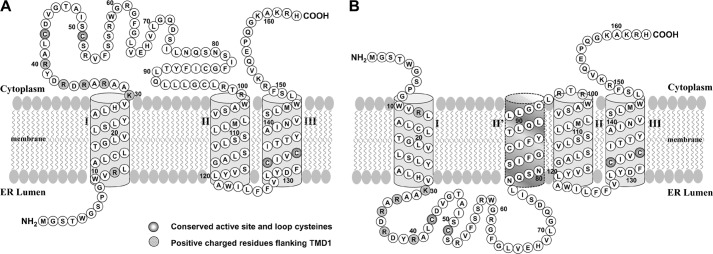
Vitamin K epoxide reductase (VKOR) is essential for the production of reduced vitamin K that is required for modification of vitamin K-dependent proteins. Three- and four-transmembrane domain (TMD) topology models have been proposed for VKOR. They are based on in vitro glycosylation mapping of the human enzyme and the crystal structure of a bacterial (Synechococcus) homologue, respectively. These two models place the functionally disputed conserved loop cysteines, Cys-43 and Cys-51, on different sides of the endoplasmic reticulum (ER) membrane. In this study, we fused green fluorescent protein to the N or C terminus of human VKOR, expressed these fusions in HEK293 cells, and examined their topologies by fluorescence protease protection assays. Our results show that the N terminus of VKOR resides in the ER lumen, whereas its C terminus is in the cytoplasm. Selective modification of cysteines by polyethylene glycol maleimide confirms the cytoplasmic location of the conserved loop cysteines. Both results support a three-TMD model of VKOR. Interestingly, human VKOR can be changed to a four-TMD molecule by mutating the charged residues flanking the first TMD. Cell-based activity assays show that this four-TMD molecule is fully active. Furthermore, the conserved loop cysteines, which are essential for intramolecular electron transfer in the bacterial VKOR homologue, are not required for human VKOR whether they are located in the cytoplasm (three-TMD molecule) or the ER lumen (four-TMD molecule). Our results confirm that human VKOR is a three-TMD protein. Moreover, the conserved loop cysteines apparently play different roles in human VKOR and in its bacterial homologues.
Figures


Similar articles
-
Membrane topology and mutational analysis of Mycobacterium tuberculosis VKOR, a protein involved in disulfide bond formation and a homologue of human vitamin K epoxide reductase.Antioxid Redox Signal. 2011 Apr 15;14(8):1413-20. doi: 10.1089/ars.2010.3558. Epub 2011 Feb 18. Antioxid Redox Signal. 2011. PMID: 20969481 Free PMC article.
-
Vitamin K epoxide reductase prefers ER membrane-anchored thioredoxin-like redox partners.Proc Natl Acad Sci U S A. 2010 Aug 24;107(34):15027-32. doi: 10.1073/pnas.1009972107. Epub 2010 Aug 9. Proc Natl Acad Sci U S A. 2010. PMID: 20696932 Free PMC article.
-
The membrane topology of vitamin K epoxide reductase is conserved between human isoforms and the bacterial enzyme.Biochem J. 2016 Apr 1;473(7):851-8. doi: 10.1042/BJ20151223. Epub 2016 Jan 15. Biochem J. 2016. PMID: 26772871
-
Structure and function of vitamin K epoxide reductase.Vitam Horm. 2008;78:103-30. doi: 10.1016/S0083-6729(07)00006-4. Vitam Horm. 2008. PMID: 18374192 Review.
-
Vitamin K epoxide reductase complex subunit 1 (VKORC1): the key protein of the vitamin K cycle.Antioxid Redox Signal. 2006 Mar-Apr;8(3-4):347-53. doi: 10.1089/ars.2006.8.347. Antioxid Redox Signal. 2006. PMID: 16677080 Review.
Cited by
-
The oxidative protein folding machinery in plant cells.Protoplasma. 2013 Aug;250(4):799-816. doi: 10.1007/s00709-012-0463-x. Epub 2012 Oct 23. Protoplasma. 2013. PMID: 23090240 Review.
-
Evaluation of oral anticoagulants with vitamin K epoxide reductase in its native milieu.Blood. 2018 Nov 1;132(18):1974-1984. doi: 10.1182/blood-2018-05-846592. Epub 2018 Aug 8. Blood. 2018. PMID: 30089628 Free PMC article.
-
The catalytic mechanism of vitamin K epoxide reduction in a cellular environment.J Biol Chem. 2021 Jan-Jun;296:100145. doi: 10.1074/jbc.RA120.015401. Epub 2020 Dec 10. J Biol Chem. 2021. PMID: 33273012 Free PMC article.
-
Disulfide bond formation in prokaryotes: history, diversity and design.Biochim Biophys Acta. 2014 Aug;1844(8):1402-14. doi: 10.1016/j.bbapap.2014.02.014. Epub 2014 Feb 25. Biochim Biophys Acta. 2014. PMID: 24576574 Free PMC article. Review.
-
Functional Study of the Vitamin K Cycle Enzymes in Live Cells.Methods Enzymol. 2017;584:349-394. doi: 10.1016/bs.mie.2016.10.015. Epub 2016 Nov 22. Methods Enzymol. 2017. PMID: 28065270 Free PMC article.
References
-
- Rost S., Fregin A., Ivaskevicius V., Conzelmann E., Hörtnagel K., Pelz H. J., Lappegard K., Seifried E., Scharrer I., Tuddenham E. G., Müller C. R., Strom T. M., Oldenburg J. (2004) Mutations in VKORC1 cause warfarin resistance and multiple coagulation factor deficiency type 2. Nature 427, 537–541 - PubMed
-
- Li T., Chang C. Y., Jin D. Y., Lin P. J., Khvorova A., Stafford D. W. (2004) Identification of the gene for vitamin K epoxide reductase. Nature 427, 541–544 - PubMed
-
- Kamali F., Wynne H. (2010) Pharmacogenetics of warfarin. Annu. Rev. Med. 61, 63–75 - PubMed
-
- Stafford D. W. (2005) The vitamin K cycle. J. Thromb. Haemost. 3, 1873–1878 - PubMed
Publication types
MeSH terms
Substances
Grants and funding
LinkOut - more resources
Full Text Sources
Other Literature Sources
Molecular Biology Databases
