. 2012 Oct 26;287(44):37185-94.
doi: 10.1074/jbc.M112.404400. Epub 2012 Aug 24.

Francisella DnaK inhibits tissue-nonspecific alkaline phosphatase

Affiliations

Francisella DnaK inhibits tissue-nonspecific alkaline phosphatase

Bernard P Arulanandam et al. J Biol Chem. .

Abstract

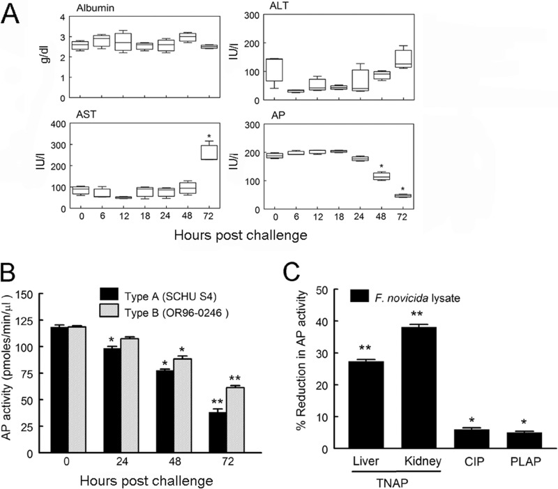
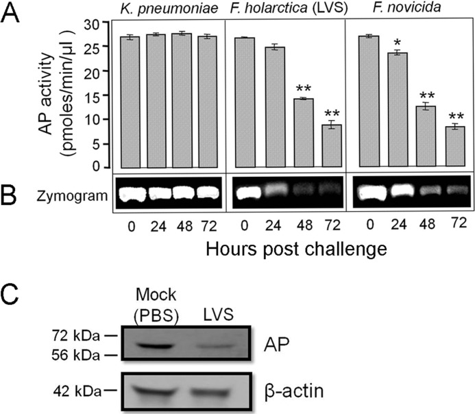
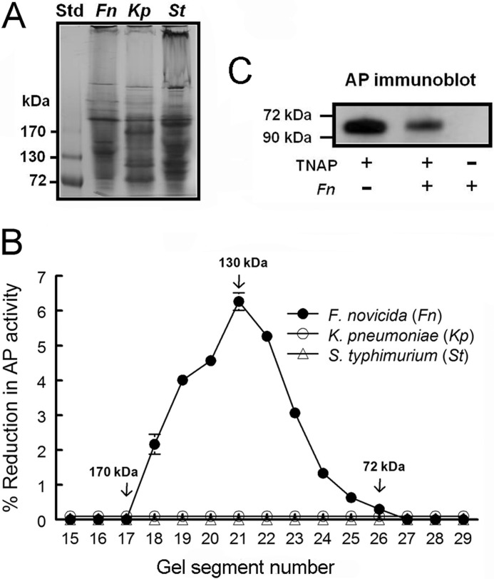
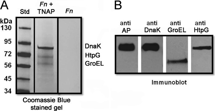
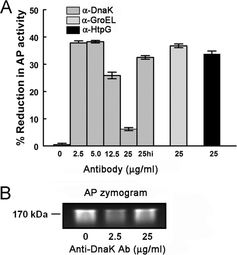
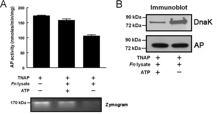
Following pulmonary infection with Francisella tularensis, we observed an unexpected but significant reduction of alkaline phosphatase, an enzyme normally up-regulated following inflammation. However, no reduction was observed in mice infected with a closely related gram-negative pneumonic organism (Klebsiella pneumoniae) suggesting the inhibition may be Francisella-specific. In similar fashion to in vivo observations, addition of Francisella lysate to exogenous alkaline phosphatase (tissue-nonspecific isozyme) was inhibitory. Partial purification and subsequent proteomic analysis indicated the inhibitory factor to be the heat shock protein DnaK. Incubation with increasing amounts of anti-DnaK antibody reduced the inhibitory effect in a dose-dependent manner. Furthermore, DnaK contains an adenosine triphosphate binding domain at its N terminus, and addition of adenosine triphosphate enhances dissociation of DnaK with its target protein, e.g. alkaline phosphatase. Addition of adenosine triphosphate resulted in decreased DnaK co-immunoprecipitated with alkaline phosphatase as well as reduction of Francisella-mediated alkaline phosphatase inhibition further supporting the binding of Francisella DnaK to alkaline phosphatase. Release of DnaK via secretion and/or bacterial cell lysis into the extracellular milieu and inhibition of plasma alkaline phosphatase could promote an orchestrated, inflammatory response advantageous to Francisella.


Figures

FIGURE 1.
FIGURE 1.
Inhibition of murine alkaline phosphatase activity by Francisella spp. A, BALB/c mice (three per group) were challenged i.n. with 400 cfu of F. novicida. Mice were bled at the indicated time points, and plasma albumin content, transaminase (ALT and AST), and AP activities were determined using an Olympus AU640e Chemistry Immuno Analyzer. Enzyme activity was reported as IU/liter (using PNPP as substrate) and albumin content g/dl. Mean values ± S.D. are shown for all experiments. Significant differences in enzymatic activities between mice prior to (0 h) and post-F. novicida challenge (48 and 72 h) are indicated (*, p < 0.05, Student's t test). Results are representative of three independent experiments. B, BALB/c mice (3–5 per group) were challenged i.n. with either 100 cfu of type A or B Francisella. Mice were bled at 24, 48, and 72 h post-challenge. Plasma was prepared and assayed using PNPP substrate as described previously under “Experimental Procedures.” Mean values ± S.D. are shown for all experiments. Significant differences in plasma AP activities between mice prior to and post-bacterial challenge are indicated (*, p < 0.05; **, p < 0.01). C, inhibitory effect of Francisella lysate supernatant material on liver and kidney TNAP, calf intestinal AP (CIP), and placental AP (PLAP) isozymes (equivalent units) was determined in triplicate as described previously under “Experimental Procedures” using 4-MUP as substrate. Mean values ± S.D. are shown for all experiments. Significant differences in AP activities are indicated (*, p < 0.05; **, p < 0.01).
FIGURE 2.
FIGURE 2.
Specificity of inhibition of plasma alkaline phosphatase. BALB/c mice (3–5 per group) were challenged i.n. with 400 cfu of either K. pneumoniae, F. holartica (LVS), or F. novicida. Mice were bled prior to challenge (0 h) and at 24, 48, and 72 h post-challenge. A, plasma AP activity was measured spectrophotometrically with PNPP substrate and reported as pmol/min/μl plasma. Mean values ± S.D. are shown for all experiments. Significant differences in plasma AP activities are indicated (*, p < 0.05; **, p < 0.01). B, respective plasma samples were subjected to PAGE, and AP activity was visualized under UV light using 4-MUP substrate as described previously under “Experimental Procedures.” C, representative Western blot analysis of PBS mock-treated and LVS-infected plasma (72 h) using anti-AP antibody. β-Actin detected by an anti-actin antibody was used as a protein loading control (42 kDa).
FIGURE 3.
FIGURE 3.
Fractionation of F. novicida inhibitory factor(s) by PAGE. A, bacterial lysate protein (100 μg) obtained from early stationary phase cultures of F. novicida (Fn), K. pneumonia (Kp), and S. typhimurium (St) along with molecular weight standards (Std) were separated on a 4–15% gradient gel and stained with Coomassie Blue. B, similarly prepared gel but without staining was cut in 2-mm segments (numbered from top to bottom). Protein was eluted from the respective gel segments and incubated with TNAP for 4 h, and hydrolysis of 4-MUP was carried out as described previously under “Experimental Procedures.” Reduction of total TNAP activity per gel segment was calculated as follows: TNAP + respective eluate/TNAP control × 100. C, reduction of TNAP protein following incubation (4 h) with total Francisella lysate (Fn) was visualized by Western blot analysis using an anti-AP antibody as described previously under “Experimental Procedures.”
FIGURE 4.
FIGURE 4.
Fractionation of F. novicida inhibitory factor(s) by DEAE-anion exchange chromatography. A, total F. novicida lysate was loaded onto a DEAE-anion exchange column and fractionated as described previously under “Experimental Procedures.” Bound protein was eluted using increasing concentrations of NaCl (50, 100, 150, and 200 mm). Inhibition of AP by each NaCl eluate was measured following a 4-h incubation with TNAP as described previously under “Experimental Procedures.” B, electrophoretic analysis of fractionated (Coomassie Blue-stained) Francisella lysate was carried out as described previously under “Experimental Procedures.” Unfractionated Francisella lysate is represented by Fn. Unbound protein, i.e. column breakthrough, is represented by BT.
FIGURE 5.
FIGURE 5.
F. novicida DnaK, GroEL, and HtpG proteins were co-immunoprecipitated with TNAP using anti-TNAP antibody-coupled beads. F. novicida (Fn) lysate (360 μl, 500 μg of protein) mixed with TNAP (40 μl, 142 μg) or F. novicida lysate alone were incubated with anti-TNAP antibody coupled with AminoLink Coupling Resin for 6–8 h at 5 °C. Co-immunoprecipitated proteins were separated on 4–15% SDS-polyacrylamide gels and visualized either by Coomassie Blue staining (A) or Western blotting (B) using anti-AP, anti-DnaK, anti-GroEL, or anti-HtpG antibodies as described under “Experimental Procedures.”
FIGURE 6.
FIGURE 6.
F. novicida-mediated AP inhibition was abrogated by anti-DnaK antibody. A, F. novicida cell lysate was incubated with TNAP in the absence (0) or presence of increasing concentrations (2.5 to 25 μg/ml as indicated) of anti-DnaK, anti-GroEL (25 μg/ml), anti-HtpG (25 μg/ml), or 25 μg/ml heat-inactivated anti-DnaK (25hi) antibodies for 4 h. Inhibition of AP was determined for each antibody treatment as described previously under “Experimental Procedures.” B, zymogram analysis of AP activity in the absence (0) or presence of anti-DnaK antibody (Ab) (2.5 and 25 μg/ml).
FIGURE 7.
FIGURE 7.
Exogenous ATP-reduced F. novicida-mediated AP inhibition. A, F. novicida (Fn) cell lysate was incubated with TNAP in the absence or presence of 4 mm ATP-MgSO4 for 4 h. Incubation of TNAP alone for 4 h was used as control. AP activity was measured using 4-MUP substrate as described previously under “Experimental Procedures” and reported as nmol/min/mg. Shown below the inhibition profile is the zymogram analysis of TNAP ± ATP. B, anti-TNAP antibody coupled with AminoLink coupling resin was mixed with F. novicida cell lysate material (500 μg of protein) and TNAP (142 μg) in the presence and/or absence of 4 mm ATP for 6–8 h at 5 °C. Proteins captured by anti-TNAP antibody-coupled resin were separated on a 4–15% SDS-polyacrylamide gel, and DnaK co-immunoprecipitation with TNAP was analyzed by Western blotting using anti-DnaK and anti-AP antibodies as described under “Experimental Procedures.”

Similar articles

References

    1. Ellis J., Oyston P. C., Green M., Titball R. W. (2002) Tularemia. Clin. Microbiol. Rev. 15, 631–646 - PMC - PubMed
    1. Tärnvik A. (1989) Nature of protective immunity to Francisella tularensis. Rev. Infect. Dis. 11, 440–451 - PubMed
    1. Chong A., Wehrly T. D., Nair V., Fischer E. R., Barker J. R., Klose K. E., Celli J. (2008) The early phagosomal stage of Francisella tularensis determines optimal phagosomal escape and Francisella pathogenicity island protein expression. Infect. Immun. 76, 5488–5499 - PMC - PubMed
    1. Cong Y., Yu J. J., Guentzel M. N., Berton M. T., Seshu J., Klose K. E., Arulanandam B. P. (2009) Vaccination with a defined Francisella tularensis subsp. novicida pathogenicity island mutant (DeltaiglB) induces protective immunity against homotypic and heterotypic challenge. Vaccine 27, 5554–5561 - PMC - PubMed
    1. Conlan J. W., Shen H., Golovliov I., Zingmark C., Oyston P. C., Chen W., House R. V., Sjöstedt A. (2010) Differential ability of novel attenuated targeted deletion mutants of Francisella tularensis subspecies tularensis strain SCHU S4 to protect mice against aerosol challenge with virulent bacteria. Effects of host background and route of immunization. Vaccine 28, 1824–1831 - PMC - PubMed

Publication types

MeSH terms

LinkOut - more resources