. 2012 Oct 12;287(42):35260-35274.
doi: 10.1074/jbc.M112.398859. Epub 2012 Aug 25.

Structure/function relationships of adipose phospholipase A2 containing a cys-his-his catalytic triad

Affiliations

Structure/function relationships of adipose phospholipase A2 containing a cys-his-his catalytic triad

Xiao-Yan Pang et al. J Biol Chem. .

Abstract

Adipose phospholipase A(2) (AdPLA or Group XVI PLA(2)) plays an important role in the onset of obesity by suppressing adipose tissue lipolysis. As a consequence, AdPLA-deficient mice are resistant to obesity induced by a high fat diet or leptin deficiency. It has been proposed that AdPLA mediates its antilipolytic effects by catalyzing the release of arachidonic acid. Based on sequence homology, AdPLA is part of a small family of acyltransferases and phospholipases related to lecithin:retinol acyltransferase (LRAT). To better understand the enzymatic mechanism of AdPLA and LRAT-related proteins, we solved the crystal structure of AdPLA. Our model indicates that AdPLA bears structural similarity to proteins from the NlpC/P60 family of cysteine proteases, having its secondary structure elements configured in a circular permutation of the classic papain fold. Using both structural and biochemical evidence, we demonstrate that the enzymatic activity of AdPLA is mediated by a distinctive Cys-His-His catalytic triad and that the C-terminal transmembrane domain of AdPLA is required for the interfacial catalysis. Analysis of the enzymatic activity of AdPLA toward synthetic and natural substrates indicates that AdPLA displays PLA(1) in addition to PLA(2) activity. Thus, our results provide insight into the enzymatic mechanism and biochemical properties of AdPLA and LRAT-related proteins and lead us to propose an alternate mechanism for AdPLA in promoting adipose tissue lipolysis that is not contingent on the release of arachidonic acid and that is compatible with its combined PLA(1)/A(2) activity.


Figures

FIGURE 1.
FIGURE 1.
Phase anomalous difference map (green mesh) contoured at 4σ using diffraction data collected at λ = 1.7463 Å. The largest peaks were observed near Cys residues 89 and 113.
FIGURE 2.
FIGURE 2.
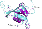
Superposition of AdPLA structures determined by NMR (Protein Data Bank code 2KYT; colored cyan) and x-ray crystallography (Protein Data Bank code 4FA0; colored magenta). The N- and C-terminal (term) ends are noted, and the disordered loop in the crystal structure is indicated by the asterisks.
FIGURE 3.
FIGURE 3.
Comparison of the NMR (Protein Data Bank code 2KYT) and x-ray structures (Protein Data Bank codes 4DOT and 4FA0) of AdPLA. A, loop regions 27–33, 71–74, 79–87, and 106–112 in AdPLA structures determined by NMR (Protein Data Bank code 2KYT; colored cyan) and x-ray crystallography (Protein Data Bank code 4FA0; colored magenta) show the largest difference between the two structures. B, loop region 104–110 contains the largest differences between the AdPLA crystal structure reported here (Protein Data Bank code 4FA0; colored magenta) and the previously reported structure (Protein Data Bank code 4DOT; colored green). C, plot of root mean square deviations (RMSD) between Cα atoms of Protein Data Bank code 2KYT versus Protein Data Bank code 4FA0 (solid gray line) and Protein Data Bank code 4DOT versus Protein Data Bank code 4FA0 (dashed black line) models of the AdPLA. D, plot of average B-factors values of Protein Data Bank code 4DOT (dashed black line) and Protein Data Bank code 4FA0 (solid gray line).
FIGURE 4.
FIGURE 4.
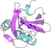
Crystal structure of AdPLA colored by secondary structure and showing the Cys113-His23-His35 catalytic triad.
FIGURE 5.
FIGURE 5.
Phospholipase A2 activity of MBP-FL-AdPLA. BODIPY PC-A2 was used to measure the rate of reaction as a function of substrate concentration. The 488/515 nm fluorescence signal of liposomes containing various concentrations of BODIPY PC-A2 substrate (structure shown in top panel) was recorded for 1 min to monitor the background emission. No significant increase in background fluorescence emission was observed. Purified MBP-FL-AdPLA protein was injected at 1 min (indicated by arrow), and the fluorescence signal at 488/515 nm was recorded for the next 4 min. A standard curve of BODIPY FL C5, the cleavage product of BODIPY PC-A2, was used for evaluating the amount of product formed by MBP-FL-AdPLA-mediated hydrolysis. Representative enzyme reaction progress curves are shown in the bottom panel. The inset shows the initial rates of reaction as a function of substrate concentration. Data represent means ± S.E. (error bars; n = 3). The enzyme kinetic data were analyzed through sum-of-squares non-linear regression to derive the Vmax and Km.
FIGURE 6.
FIGURE 6.
Deletion and mutagenesis analysis of MBP-FL-AdPLA. Top panel, rate of reaction of MBP-FL-AdPLA and MBP-T-AdPLA as a function of enzyme concentration. The rate of PLA2 activity of MBP-FL-AdPLA and MBP-T-AdPLA was measured during steady-state conditions using the fluorescent substrate BODIPY PC-A2. A standard curve of BODIPY FL C5 was used for evaluating the amount of product formed by MBP-FL-AdPLA-mediated hydrolysis. Data represent means ± S.E. (error bars; n = 3). Bottom panel, the rate of activity of wild type and active site mutants of MBP-FL-AdPLA. Equal amounts of purified recombinant proteins were examined for hydrolysis of BODIPY PC-A2. The rate of activity is expressed as a percentage of the activity of wild type protein.
FIGURE 7.
FIGURE 7.
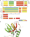
Amide hydrogen/deuterium exchange analysis of T-AdPLA, MBP-T-AdPLA, and MBP-FL-AdPLA. A, the percentage of maximal deuterium incorporation in T-AdPLA (upper ribbon), MBP-T-AdPLA (middle ribbon), and MBP-FL-AdPLA (lower ribbon) peptidic regions is shown. In each ribbon, the percentage of deuteration at each of the labeling durations is shown from 10 to 10,000 s (top to bottom). Differential coloring indicates the percentage of maximal labeling in a given time point. B, crystal structure of AdPLA colored based on fast (red) or intermediate (green) hydrogen/deuterium exchange rates. The N terminus and the residues His23, His35, and Cys113 composing the catalytic triad are indicated. Dotted lines represent the C-terminal strands and the 35–59 loop region for which there are no x-ray data.
FIGURE 8.
FIGURE 8.
Analysis of the specificity of the phospholipase activity of MBP-FL-AdPLA and MBP-T-AdPLA. PLA1/PLA2 activity was assayed using liposome compositions containing an equimolar mixture of 18:1-18:1 PG and one of five different PC species examined as indicated at the top of each panel. The liposomes were incubated for 15 min with either MBP-FL-AdPLA, MBP-T-AdPLA, buffer control (labeled no enzyme), bee venom PLA2, or PLA1 from T. lanuginosus (C). The resulting fatty acids were extracted and analyzed by HPLC-charged aerosol detection. The fatty acid products of reactions carried out (solid black line chromatograms) were identified based on the elution profile in comparison with authentic standards (dashed gray line chromatograms). The oleate (18:1) fatty acid product of the hydrolysis of 18:1-18:1 PG (A–F) or 18:1-18:1 PC (F) by MBP-FL-AdPLA, bee venom PLA2, or PLA1 from T. lanuginosus is indicated by a black star (*). The experiment was conducted in triplicate and repeated.

References

    1. Coppack S. W., Jensen M. D., Miles J. M. (1994) In vivo regulation of lipolysis in humans. J. Lipid Res. 35, 177–193 - PubMed
    1. Villena J. A., Roy S., Sarkadi-Nagy E., Kim K. H., Sul H. S. (2004) Desnutrin, an adipocyte gene encoding a novel patatin domain-containing protein, is induced by fasting and glucocorticoids: ectopic expression of desnutrin increases triglyceride hydrolysis. J. Biol. Chem. 279, 47066–47075 - PubMed
    1. Zimmermann R., Strauss J. G., Haemmerle G., Schoiswohl G., Birner-Gruenberger R., Riederer M., Lass A., Neuberger G., Eisenhaber F., Hermetter A., Zechner R. (2004) Fat mobilization in adipose tissue is promoted by adipose triglyceride lipase. Science 306, 1383–1386 - PubMed
    1. Jenkins C. M., Mancuso D. J., Yan W., Sims H. F., Gibson B., Gross R. W. (2004) Identification, cloning, expression, and purification of three novel human calcium-independent phospholipase A2 family members possessing triacylglycerol lipase and acylglycerol transacylase activities. J. Biol. Chem. 279, 48968–48975 - PubMed
    1. Osuga J., Ishibashi S., Oka T., Yagyu H., Tozawa R., Fujimoto A., Shionoiri F., Yahagi N., Kraemer F. B., Tsutsumi O., Yamada N. (2000) Targeted disruption of hormone-sensitive lipase results in male sterility and adipocyte hypertrophy, but not in obesity. Proc. Natl. Acad. Sci. U.S.A. 97, 787–792 - PMC - PubMed

Publication types

MeSH terms

Substances

Associated data