. 2012 Oct 15;303(8):L711-9.
doi: 10.1152/ajplung.00037.2012. Epub 2012 Aug 24.

Oxygen dose responsiveness of human fetal airway smooth muscle cells

Affiliations

Oxygen dose responsiveness of human fetal airway smooth muscle cells

William R Hartman et al. Am J Physiol Lung Cell Mol Physiol. .

Abstract

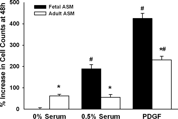
Maintenance of blood oxygen saturation dictates supplemental oxygen administration to premature infants, but hyperoxia predisposes survivors to respiratory diseases such as asthma. Although much research has focused on oxygen effects on alveoli in the setting of bronchopulmonary dysplasia, the mechanisms by which oxygen affects airway structure or function relevant to asthma are still under investigation. We used isolated human fetal airway smooth muscle (fASM) cells from 18-20 postconceptual age lungs (canalicular stage) to examine oxygen effects on intracellular Ca(2+) ([Ca(2+)](i)) and cellular proliferation. fASM cells expressed substantial smooth muscle actin and myosin and several Ca(2+) regulatory proteins but not fibroblast or epithelial markers, profiles qualitatively comparable to adult human ASM. Fluorescence Ca(2+) imaging showed robust [Ca(2+)](i) responses to 1 μM acetylcholine (ACh) and 10 μM histamine (albeit smaller and slower than adult ASM), partly sensitive to zero extracellular Ca(2+). Compared with adult, fASM showed greater baseline proliferation. Based on this validation, we assessed fASM responses to 10% hypoxia through 90% hyperoxia and found enhanced proliferation at <60% oxygen but increased apoptosis at >60%, effects accompanied by appropriate changes in proliferative vs. apoptotic markers and enhanced mitochondrial fission at >60% oxygen. [Ca(2+)](i) responses to ACh were enhanced for <60% but blunted at >60% oxygen. These results suggest that hyperoxia has dose-dependent effects on structure and function of developing ASM, which could have consequences for airway diseases of childhood. Thus detrimental effects on ASM should be an additional consideration in assessing risks of supplemental oxygen in prematurity.


Figures

Fig. 1.
Fig. 1.
Serum-deprived human fetal airway smooth muscle (fASM) cells display mononucleated spindle-shaped morphology typical of smooth muscle cells. Scale bar = 50 μm for left and 10 μm for right.
Fig. 2.
Fig. 2.
Expression of intracellular Ca2+ ([Ca2+]i) regulatory proteins and smooth muscle markers in fASM cells. Western analysis of whole cell lysates showed substantial expression of contractile proteins (smooth muscle actin and myosin heavy chain, MHC), receptors for bronchoconstrictor agonists (M3 muscarinic receptor; acetylcholine receptor, AChR); H1 histaminergic receptor; neurokinin receptors (NK1R and NK2R) as well a number of [Ca2+]i regulatory mechanisms including sarcoendoplasmic reticulum Ca2+ ATPase (SERCA) and IP3 receptor (IP3R), and to lesser extents ryanodine receptors (RyR), transient receptor potential channel TRPC3, and store-operated Ca2+ entry regulator STIM1. In contrast, markers of epithelial cells (E-cadherin) or fibroblasts (fibroblast surface protein; FSP) were absent. Values (means ± SE) from n of 5 samples normalized to GAPDH in bar graph. *Significant difference between fASM and adult ASM cells (P < 0.05).
Fig. 3.
Fig. 3.
Expression of caveolin-1 in fASM cells. Compared with adult ASM cells, fASM cells expressed significantly greater amounts of the constituent caveolar protein caveolin-1. Exposure to the cholesterol-chelating agent methyl-β-cyclodextrin (CD) significantly reduced caveolin-1 expression, verifying membrane expression of this protein. Values (means ± SE) from n of 5 samples normalized to GAPDH in bar graph. *Significant CD effect; #significant difference between fASM and adult ASM cells (P < 0.05).
Fig. 4.
Fig. 4.
[Ca2+]i responses of fASM cells to agonists. Exposure of fASM cells to 1 μM ACh or 10 μM histamine induced a large peak [Ca2+]i response followed by a lower plateau level above baseline (A). HBSS, Hanks's Balanced Salt Solution. Overnight exposure to the neurokinin receptor agonist Substance P (SP; 1 μM) resulted in enhanced [Ca2+]i responses to subsequent 1 μM ACh exposure (B). In general, compared with adult ASM cells that also produced qualitatively similar responses, the peak [Ca2+]i responses to agonist were significantly smaller in fASM cells for ACh (with or without SP) and histamine. Values (means ± SE) from n of 5 samples in bar graph (C). *Significant difference between fASM and adult ASM cells; #significant SP effect (P < 0.05).
Fig. 5.
Fig. 5.
Mechanisms of [Ca2+]i responses in fASM cells. Removal of extracellular Ca2+ significantly blunted the [Ca2+]i responses to ACh (shown) (A), confirming a component of Ca2+ influx. The influx component, relative to overall [Ca2+]i response, was larger in fASM cells compared with adult ASM cells. Conversely, fASM cells showed a transient [Ca2+]i response to 5 mM caffeine (albeit slower and smaller than adult ASM), indicating the presence of SR Ca2+ release and functional RyR channels (B). Separately, exposure to 10 mM of the plasma membrane cholesterol-chelating agent methyl-β-cyclodextrin (CD) for 1 h decreased baseline [Ca2+]i and furthermore blunted [Ca2+]i responses to ACh, indicating a role for plasma membrane caveolae in [Ca2+]i regulation of fASM cells (C). Values (means ± SE) from n of 5 samples in bar graph (D). *Significant difference between fASM and adult ASM cells (P < 0.05). #Significant effect of 0 Ca2+ HBSS or CD.
Fig. 6.
Fig. 6.
Proliferation of fASM cells. Cellular proliferation was determined using a CyQuant assay. In serum-depleted media, fASM cells showed minimal proliferation at 48 h. The presence of 0.5% serum substantially enhanced proliferation, especially in fASM cells, as did the standard mitogen platelet-derived growth factor (PDGF). Values (means ± SE) from n of 5 samples. Percent increase from initial cell count of 5,000. *Significant difference between fASM and adult ASM cells (P < 0.05). #Significant effect of serum or PDGF.
Fig. 7.
Fig. 7.
Proliferation responses of fASM cells to oxygen. Cellular proliferation was determined using a fluorescent CyQuant assay. Compared with normoxic controls, fASM cells showed enhanced proliferation in 10% hypoxia at 48 h. However, with increasing oxygen concentration beyond normoxia, cell proliferation progressively increased until 50%, beyond which proliferation quickly dropped off to below normoxic levels, suggesting increasing cell death. Values (means ± SE) from n of 5 samples. Percent increase from initial cell count of 5,000. *Significant difference in cell number from normoxia (P < 0.05).
Fig. 8.
Fig. 8.
Effect of oxygen on expression of proliferation and apoptosis proteins in fASM cells. A: Western analysis of whole cell lysates showed increased expression for markers of cellular proliferation (proliferating cell nuclear antigen, PCNA, Cyclin E) when fASM cells were exposed for 48 h to 10% hypoxia or to moderate levels of hyperoxia until 40%. Correspondingly, antiapoptotic proteins such as BCL-2 were higher. However, beyond 40%, expression of proapoptotic proteins such as p27Kip1, caspase 9, and cytochrome c progressively increased with increasing oxygen concentration. Values (means ± SE) from n of 5 samples normalized to GAPDH in bar graph (B). *Significant difference from normoxia (P < 0.05).
Fig. 9.
Fig. 9.
Mitochondrial fragmentation in fASM cells. A: fluorescence imaging of MitoTracker-labeled fASM cells in normoxia showed reticular mitochondrial patterns. With increasing oxygen concentrations, mitochondria took on a more punctate and aggregated appearance with short branching patterns such that at 80% oxygen, mostly aggregated mitochondria were observed. B: mitochondrial fusion protein (Mfn2) expression was significantly decreased at higher oxygen tensions, whereas the mitochondrial fission protein Drp1 was decreased. Values (means ± SE) from n of 5 samples normalized to GAPDH in bar graph. *Significant difference from normoxia (P < 0.05).

Similar articles

Cited by

References

    1. Agani FH, Kuo NT, Chang CH, Dreshaj IA, Farver CF, Krause JE, Ernsberger P, Haxhiu MA, Martin RJ. Effect of hyperoxia on substance P expression and airway reactivity in the developing lung. Am J Physiol Lung Cell Mol Physiol 273: L40–L45, 1997 - PubMed
    1. Aravamudan B, Thompson M, Pabelick C, Prakash YS. Brain-derived neurotropic factor induces proliferation of human airway smooth muscle cells. J Cell Mol Med 16: 812–823, 2012 - PMC - PubMed
    1. Badri KR, Zhou Y, Schuger L. Embryological origin of airway smooth muscle. Proc Am Thorac Soc 5: 4–10, 2008 - PMC - PubMed
    1. Bates JH, Rincon M, Irvin CG. Animal models of asthma. Am J Physiol Lung Cell Mol Physiol 297: L401–L410, 2009 - PMC - PubMed
    1. Belik J, Jankov RP, Pan J, Yi M, Chaudhry I, Tanswell AK. Chronic O2 exposure in the newborn rat results in decreased pulmonary arterial nitric oxide release and altered smooth muscle response to isoprostane. J Appl Physiol 96: 725–730, 2004 - PubMed

Publication types

MeSH terms

LinkOut - more resources