. 2012 Sep 25;109(39):E2595-604.
doi: 10.1073/pnas.1212834109. Epub 2012 Aug 23.

ECM microenvironment regulates collective migration and local dissemination in normal and malignant mammary epithelium

Affiliations

ECM microenvironment regulates collective migration and local dissemination in normal and malignant mammary epithelium

Kim-Vy Nguyen-Ngoc et al. Proc Natl Acad Sci U S A. .

Abstract

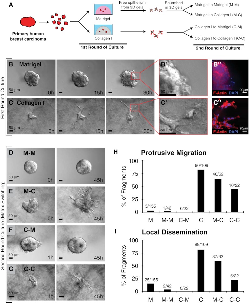
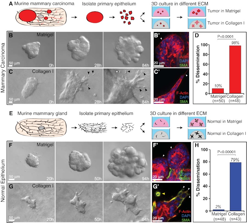
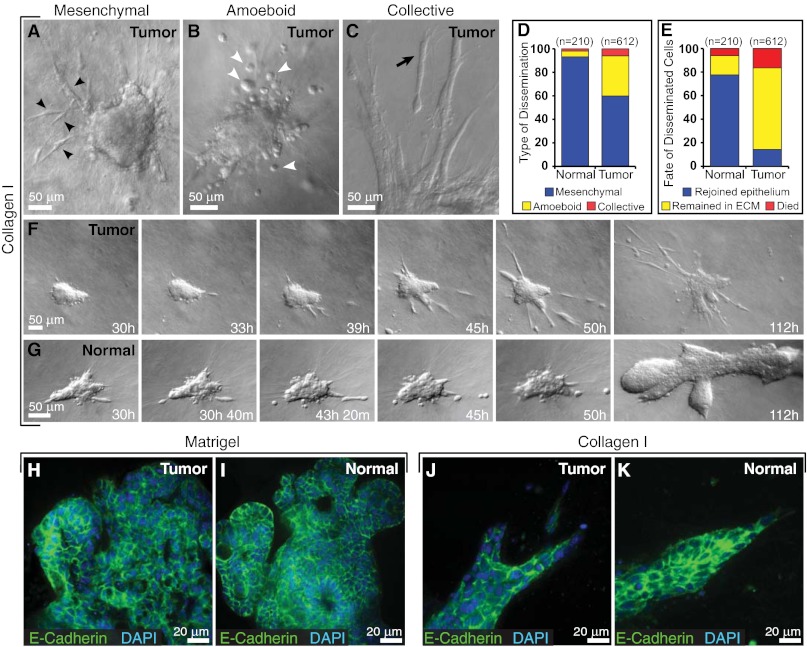
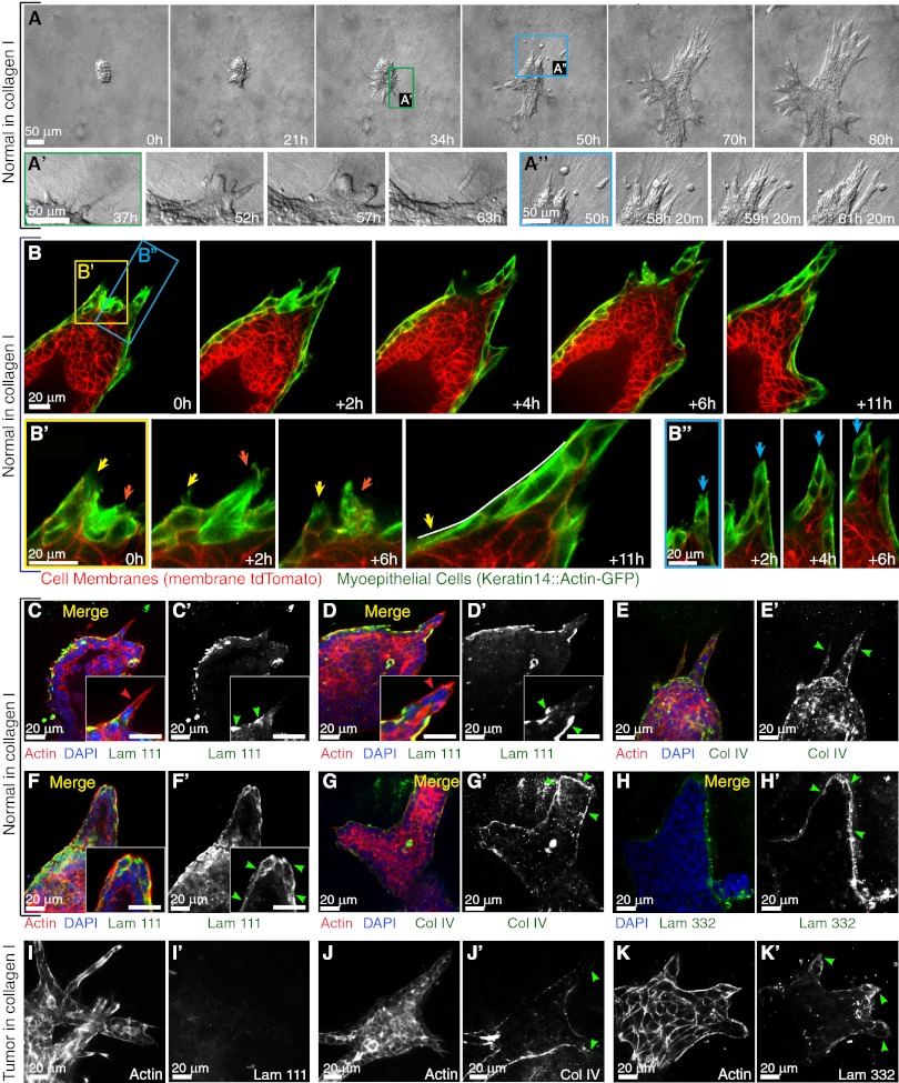
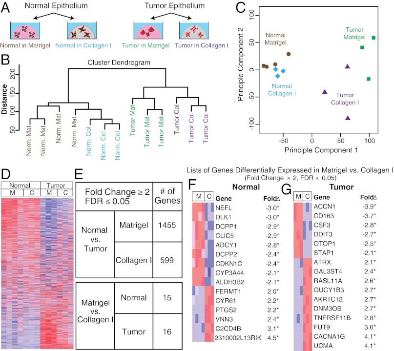
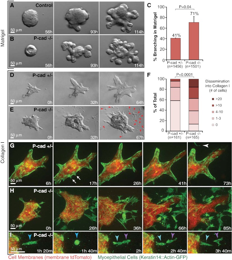
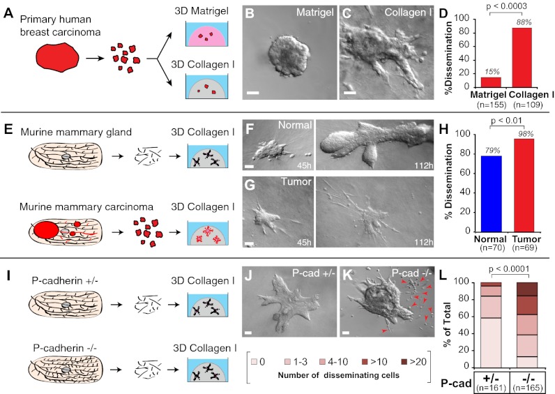
Breast cancer progression involves genetic changes and changes in the extracellular matrix (ECM). To test the importance of the ECM in tumor cell dissemination, we cultured epithelium from primary human breast carcinomas in different ECM gels. We used basement membrane gels to model the normal microenvironment and collagen I to model the stromal ECM. In basement membrane gels, malignant epithelium either was indolent or grew collectively, without protrusions. In collagen I, epithelium from the same tumor invaded with protrusions and disseminated cells. Importantly, collagen I induced a similar initial response of protrusions and dissemination in both normal and malignant mammary epithelium. However, dissemination of normal cells into collagen I was transient and ceased as laminin 111 localized to the basal surface, whereas dissemination of carcinoma cells was sustained throughout culture, and laminin 111 was not detected. Despite the large impact of ECM on migration strategy, transcriptome analysis of our 3D cultures revealed few ECM-dependent changes in RNA expression. However, we observed many differences between normal and malignant epithelium, including reduced expression of cell-adhesion genes in tumors. Therefore, we tested whether deletion of an adhesion gene could induce sustained dissemination of nontransformed cells into collagen I. We found that deletion of P-cadherin was sufficient for sustained dissemination, but exclusively into collagen I. Our data reveal that metastatic tumors preferentially disseminate in specific ECM microenvironments. Furthermore, these data suggest that breaks in the basement membrane could induce invasion and dissemination via the resulting direct contact between cancer cells and collagen I.


Conflict of interest statement

The authors declare no conflict of interest.

Figures

Fig. 1.
Fig. 1.
ECM microenvironments modulate the pattern of collective migration and local dissemination in human mammary carcinomas. (A) Schematic description of isolation and 3D culture of human mammary carcinoma fragments. In the first round of culture, tumor fragments were embedded in either 3D Matrigel or collagen I. In the second round of culture, the same tumor fragments were freed from the 3D gels and were re-embedded in the same or were swapped to the other microenvironment. (BC′′) Representative DIC time-lapse sequences of human mammary carcinomas in Matrigel (B) or collagen I (C). (B′ and C′) Enlarged views of B and C at 30 h showing the smooth and protrusive leading fronts, respectively. (B′′ and C′′) Micrographs of the border of the same mammary carcinoma embedded in Matrigel or collagen and stained with phalloidin–F-actin and DAPI. (DG) Representative frames of DIC time-lapse movies of human mammary carcinomas switched from Matrigel to Matrigel (M–M) (D), Matrigel to collagen I (M–C) (E), collagen I to Matrigel (C–M) (F), or collagen I to collagen I (C–C) (G) at 0 or 1 h in culture (Left) or 45 h in culture (Right). (H and I) Bar graphs showing the number of tumor fragments in each ECM condition with protrusive migration (H) or local dissemination (E) relative to the number of primary human tumor fragments analyzed in each condition.
Fig. 2.
Fig. 2.
The ECM governs the migratory pattern and disseminative behavior of both tumor and normal murine mammary epithelium. (A) Schematic description of isolation and 3D culture of murine tumor fragments. (B and C) Representative frames of DIC time-lapse movies of tumor fragments in Matrigel (B) and collagen I (C). Black arrowheads indicate disseminated cells, some of which are observed to proliferate (white arrowhead). (B′ and C′) Localization of actin, SMA, and DAPI in tumor fragments in Matrigel (B′) and collagen I (C′). White arrowheads mark the leading fronts. (D) Percent of tumor fragments showing cell dissemination in Matrigel and collagen I. n, total number of movies (four biological replicates, Student's t test, two-tailed, unequal variance). (E) Schematic description of isolation and 3D culture of normal mammary organoids. (F and G) Representative frames from DIC time-lapse movies of normal organoids in Matrigel (F) and collagen I (G). (F′ and G′) Localization of actin, SMA, and DAPI in normal organoids in Matrigel (F′) and collagen I (G′). White arrowheads mark the leading fronts. Yellow arrowhead indicates myoepithelial cell dissemination. (H) Percent of normal organoids showing dissemination in Matrigel and collagen I. n, total number of movies (four biological replicates, Student's t test, two-tailed, unequal variance).
Fig. 3.
Fig. 3.
Cell dissemination into collagen I is persistent in tumor and transient in normal epithelium. (AC) Tumor cells disseminate with mesenchymal (black arrowheads) (A), amoeboid (white arrowheads) (B), and collective (black arrow) (C) morphologies. (D and E) Distribution of morphological types of dissemination (D) and fate of disseminated cells in normal and tumor organoids (E) in collagen I. n, total number of disseminated cells observed in each condition. (F and G) Representative frames from DIC time-lapse movies of tumor (F) and normal organoids (G) in collagen I. (H and I) Localization of E-cadherin and DAPI in tumor (H) and normal organoids (I) cultured in Matrigel. (J and K) Localization of E-cadherin and DAPI in tumor (J) and normal organoids (K) cultured in collagen I.
Fig. 4.
Fig. 4.
Normal epithelium transiently protrudes and disseminates into collagen I but reestablishes a complete basement membrane. (A) Representative frames of a DIC time-lapse movie of a normal organoid grown in collagen I. (A′) Higher magnification of a transition from a protrusive to a smooth border with ECM. (A′′) Higher magnification of the reintegration of disseminated cells with the epithelial group. (B) Representative frames from a confocal time-lapse movie of a normal organoid grown in collagen I. (B′) Higher magnification of transient protrusions within single myoepithelial cells. Arrows indicate individual protrusions, retractions, and epithelial reorganization. (B′′) Higher magnification of a multicellular extension of myoepithelial cells (blue arrows) at the leading front. (CG) Normal epithelia in collagen reform a multicomponent basement membrane. (CD′ and F and F′) Localization of actin, DAPI, and laminin 111 in a merge of all channels (C, D, and F) and in a single channel of laminin 111 (C′, D′, and F′) in a normal organoid with a single-cell protrusion (C′), a multicellular extension (D′), and a normal organoid after reorganization (F′). All the Insets highlight negative correlation of cell protrusion (C, D, and F) and laminin 111 (C′, D′, and F′) at the leading front. (E and E′ and G and G′) Localization of actin, DAPI, and collagen IV in a merge of all channels (E and G) and in a single channel of collagen IV in a normal organoid (E′ and G′) with multicellular extensions (E′) and after reorganization (G′). (H and H′) Localization of DAPI and laminin 332 in a merge of two channels (H) and a single channel of laminin 332 (H′). (IK′) Tumor epithelia display incomplete basement membrane coverage. Single channels show the localization of actin (I, J, and K), laminin 111(I′), collagen IV (J′), and laminin 332 (K′) in tumor organoids in collagen I. Red and green arrowheads indicate actin-based protrusions and signals of basement membrane components, respectively.
Fig. 5.
Fig. 5.
Tumor and normal epithelium remain transcriptionally distinct despite morphological similarities induced by the ECM. (A) Schematic description of 3D cultures of normal and tumor murine epithelial fragments in Matrigel and collagen I for mRNA expression analyses. n = at least 3 biological replicates. (B) Complete-linkage hierarchical clustering of the experimental conditions. (C) Principal component analysis of the experimental conditions. (D) Heatmap representation of the 19,693 genes included in the microarray (blue and red indicate lower and higher expression, respectively). (E) Summary of differentially expressed genes with fold changes ≥2 and FDR ≤0.05. (F and G) Genes differentially expressed based on ECM condition in normal (F) and tumor (G). C, collagen I; M, Matrigel.
Fig. 6.
Fig. 6.
Loss of P-cadherin causes precocious branching morphogenesis in Matrigel and enhanced, sustained dissemination into collagen I. (A and B) Representative frames from DIC time-lapse movies of (A) control [P-cadherin+/+ (P-cad+/+)] and (B) P-cadherin−/− (P-cad−/−) epithelium grown in parallel in Matrigel. (C) Percent of P-cad+/− and P-cad−/− organoids branching in Matrigel on day 7. n, total number of organoids counted (three biological replicates; *P = 0.04; Student’s t test, two-tailed, unequal variance). (D and E) Representative frames from DIC time-lapse movies of P-cad+/− (D) and P-cad−/− (E) epithelium grown in parallel in collagen I. Arrowheads indicate persistent cell dissemination. (F) Distribution of number of disseminated cells per organoid in P-cad+/− and P-cad−/− epithelia. n, total number of movies (three biological replicates; *P < 0.0001; upper one-sided χ2 test). (G) Representative frames from a confocal time-lapse movie of P-cad+/−, mT/mG, K14::Actin-GFP epithelium in collagen I. Arrows indicate transient, myoepithelial-led protrusions. Arrowhead indicates a single disseminated myoepithelial cell. (H) Representative frames from a confocal time-lapse movie of enhanced myoepithelial dissemination into collagen I by P-cad−/−, mT/mG, K14::Actin-GFP epithelium. (I) Proliferation of a disseminated P-cad−/− myoepithelial cell.
Fig. P1.
Fig. P1.
The ECM regulates the dissemination of mammary epithelial cells. (A) Schematic of the isolation and 3D culture of human breast tumor organoids. (B and C) Representative images of human tumor fragments in (B) Matrigel and (C) collagen I. (D) Percentage of tumor organoids showing cell dissemination in Matrigel and collagen I. (E) Schematic of isolation and 3D culture of normal and tumor murine mammary organoids in collagen I. (F and G) Representative time-lapse images of (F) normal and (G) tumor organoids in collagen I. (H) Percentage of normal and tumor organoids that disseminated cells into collagen I. (I) Schematic of organoid isolation from P-cadherin+/− and P-cadherin−/− mouse mammary glands. (J and K) Representative images of (J) P-cadherin+/− and (K) P-cadherin−/− organoids in collagen I. (L) Distribution of the number of disseminated cells per organoid in P-cadherin+/− and P-cadherin−/− epithelia. n = total number of organoids. (Scale bars, 50 μm.)

References

    1. Friedl P, Gilmour D. Collective cell migration in morphogenesis, regeneration and cancer. Nat Rev Mol Cell Biol. 2009;10:445–457. - PubMed
    1. Ewald AJ, Brenot A, Duong M, Chan BS, Werb Z. Collective epithelial migration and cell rearrangements drive mammary branching morphogenesis. Dev Cell. 2008;14:570–581. - PMC - PubMed
    1. Ewald AJ, et al. Mammary collective cell migration involves transient loss of epithelial features and individual cell migration within the epithelium. J Cell Sci. 2012;125:2638–2654. - PMC - PubMed
    1. Hanahan D, Weinberg RA. The hallmarks of cancer. Cell. 2000;100:57–70. - PubMed
    1. Friedl P, et al. Migration of coordinated cell clusters in mesenchymal and epithelial cancer explants in vitro. Cancer Res. 1995;55:4557–4560. - PubMed

Publication types

Associated data