

Evaluation and comparison of decalcification agents on the human teeth
- PMID: 22923894
- PMCID: PMC3424938
- DOI: 10.4103/0973-029X.99070
Evaluation and comparison of decalcification agents on the human teeth
Abstract
Context: In routine histopathology, decalcification of bone and teeth is often an essential and important step during tissue processing. Various decalcifying agents have been used in the past. The rate of decalcification and the effect of decalcifying agents on the tissue and its staining characteristics are two important parameters which influence the selection of decalcifying solutions. Though some agents remove the calcium ions completely and rapidly, they adversely affect the staining characteristics and may also damage the organic components. There have been very few studies which have systematically evaluated the efficacy of these agents in decalcifying dental hard tissues.
Aims: The present study was done to evaluate the rate of decalcification of six different decalcifying agents and also their effect on staining characteristics on dental hard tissues.
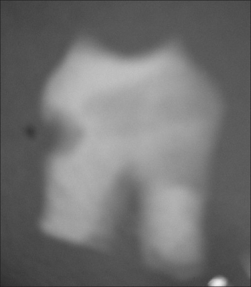
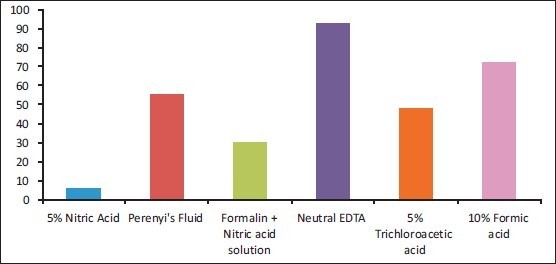
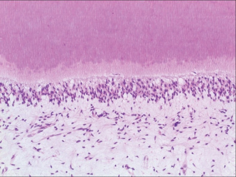
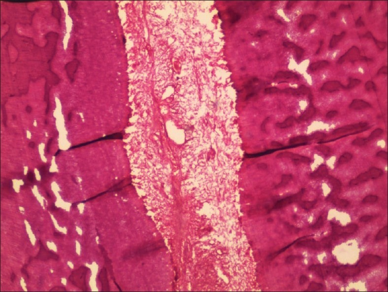
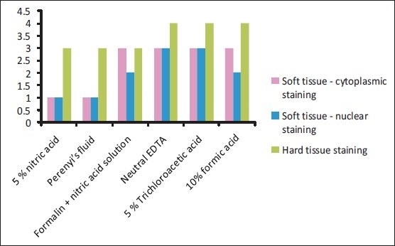
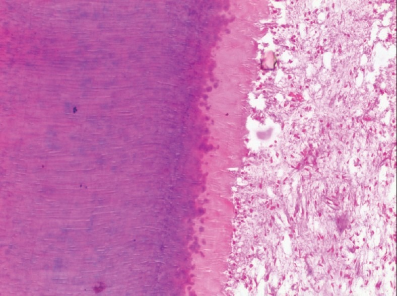
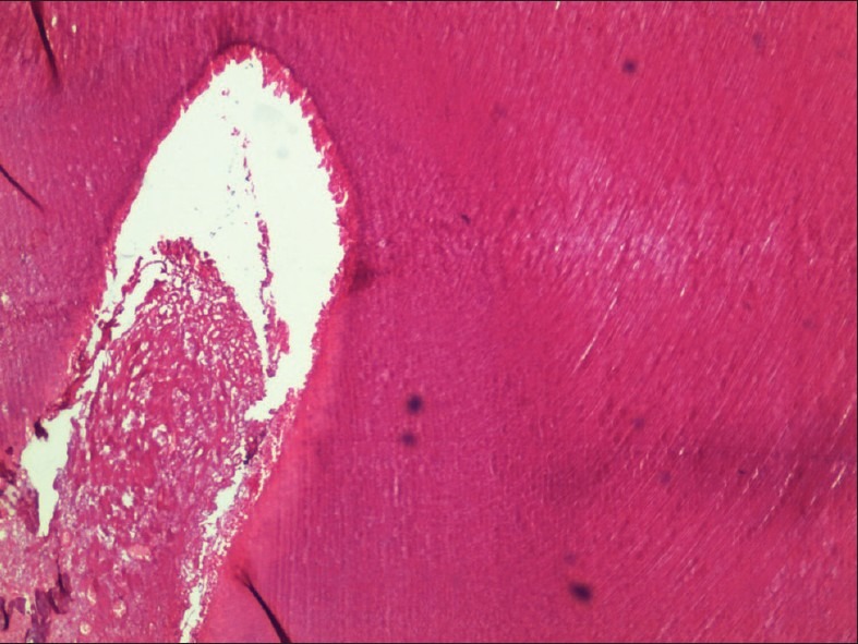
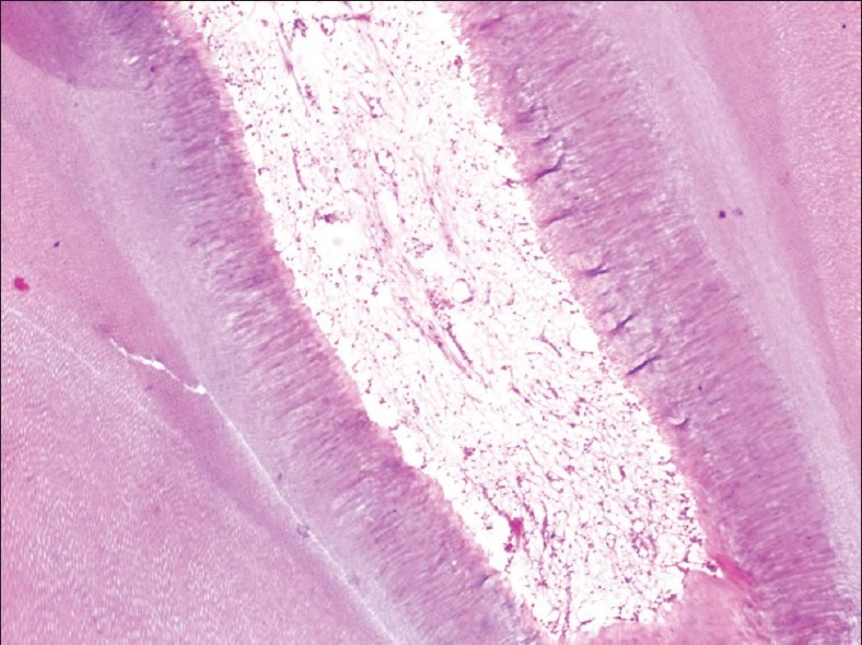
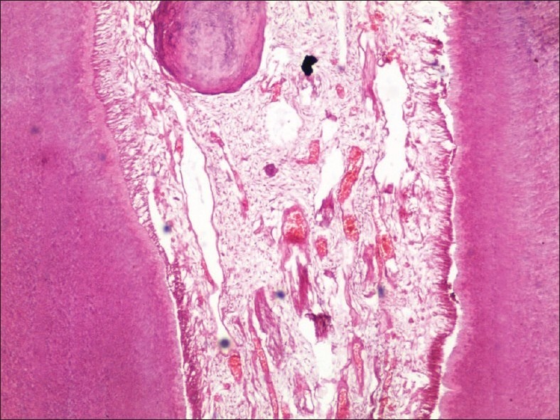
Materials and methods: Six decalcifying agents namely, neutral ethylene diamine tetra acetic acid (EDTA) decalcifying solution, 5% nitric acid, Perenyi's fluid, formalin-nitric acid, 5% trichloracetic acid, and 10% formic acid were used to decalcify 24 natural teeth (four in each solution). The endpoint of decalcification was evaluated by radiographic and chemical methods. The decalcified teeth were then routinely processed, sectioned, and stained with hematoxylin and eosin stains.
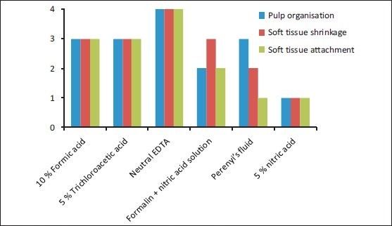
Results: Neutral EDTA was the most considerate to the soft and hard tissues and 5% nitric acid was the least considerate to the tooth structure.
Conclusions: Neutral EDTA, though being the slowest decalcifying agent among the six agents used in the study, gave excellent results for soft-tissue integrity, and best quality of both soft-tissue and hard-tissue stainings.
Keywords: 10% formic acid; 5% nitric acid; 5% trichloracetic acid; Perenyi's fluid; formalin–nitric acid; neutral EDTA decalcifying solution; pulp tissue integrity; teeth decalcification.
Conflict of interest statement
Figures
References
-
- Olympic charter published by International Olympic committee, Lausanne, Switzerland. 2011:9–21.
-
- Cook SF, Ezra-Cohn HE. A comparison of methods for decalcifying bone. J Histochem Cytochem. 1962;10:560–63.
-
- Drury RA, Wallington EA. 5th ed. Oxford: Oxford University Press; 1980. Carleton's Histological Technique; pp. 199–205.
-
- Mattuella LG, Bento LW, Vier-Pelisser FV, Araiyo FB, Fossati AC. Comparative analysis of two fixating and two decalcifying solutions for processing of human primary teeth with inactive dentin carious lesion. Rev Odonto Ciênc. 2007;22:99–105.
-
- Callis MG. Bone. In: Bancroft JD, Gamble M, editors. Theory and practice of histological techniques. 6th ed. Philadelphia: Churchill Livingstone; 2008. pp. 333–63.
LinkOut - more resources
Full Text Sources
Miscellaneous
