

Pathophysiology of Langerhans cells
- PMID: 22923897
- PMCID: PMC3424941
- DOI: 10.4103/0973-029X.99077
Pathophysiology of Langerhans cells
Abstract
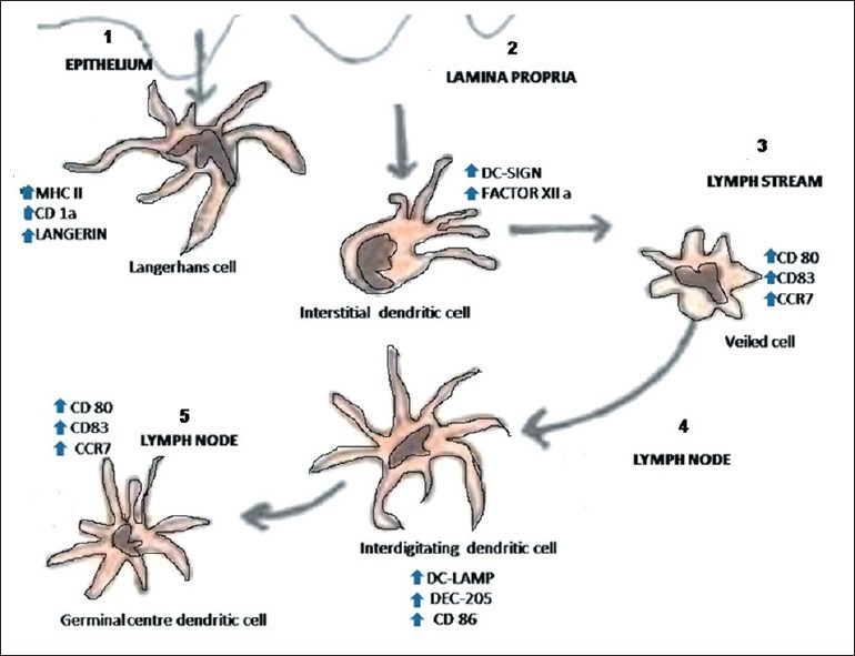
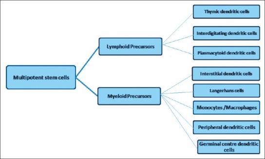
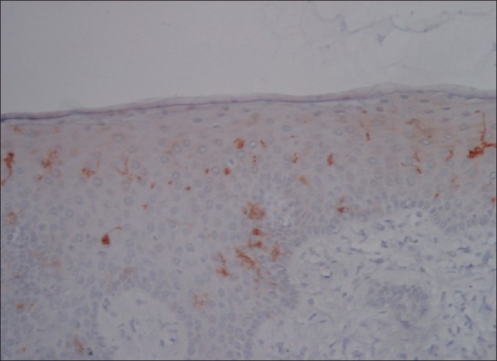
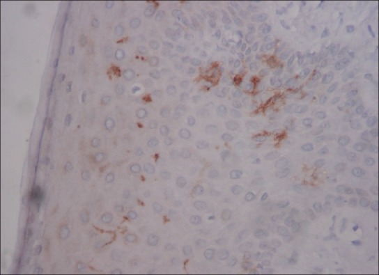
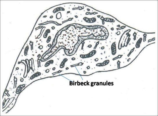
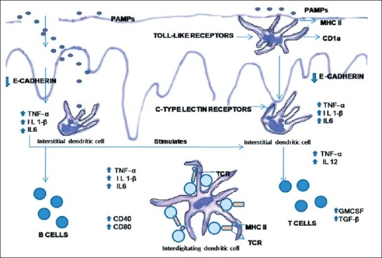
Langerhans cells (LCs) were first described by Paul Langerhans, in 1868, as dendritically shaped cells, which were located in the squamous epithelia of epidermis. Later on, these cells were identified in all stratified squamous epithelium of mammals. Dendritic cells (DCs) play an important role in local defense mechanisms in the epithelium. LCs are situated usually in the suprabasal layer of stratified squamous epithelia of oral mucosa and epidermis of skin. They constitute 3% of the cell population in epidermis. LCs are thought to act as antigen presenting cells (APCs) during initiation of immune responses. With the help of APCs, the lymphocytes are able to recognize and respond to specific microbes. In this paper we have reviewed the origin, distribution, demonstration and mechanism of action of LCs and their role in different pathological conditions.
Keywords: Antigen presenting cells; Langerhans cells; clear cells; dendritic cells.
Conflict of interest statement
Figures
References
-
- Lu FX, Jacobson RS. Oral mucosal immunity and HIV/SIV Infection. J Dent Res Mar. 2007;86:216–26. - PubMed
-
- 5th edition. Missouri: Mosby Year Book Inc; 1996. Ten Kate AR: Oral Histology. Development, structure and Function.
-
- de Witte L, Nabatov A, Pion M, Fluitsma D, de Jong MA, de Gruijl T, et al. Langerin is a natural barrier to HIV-1 transmission by Langerhans cells. Nature Med. 2007;13(3):367–71. - PubMed
-
- Lombardi T, Hauser C, Budtz-Jörgensen E. Langerhans cells: structure, function and role in oral pathological conditions. J Oral Pathol Med. 1993;22:193–202. - PubMed
LinkOut - more resources
Full Text Sources
Other Literature Sources
Miscellaneous
