

Intraosseous schwannoma of the mandible
- PMID: 22923909
- PMCID: PMC3424953
- DOI: 10.4103/0973-029X.99094
Intraosseous schwannoma of the mandible
Abstract
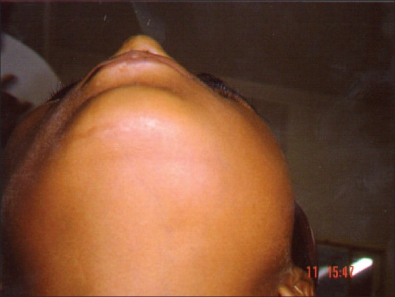
Schwannomas (neurilemmomas) are slow-growing, benign neoplasms derived from schwann cells, the sheath cells that cover myelinated nerve fibers. These tumors most commonly arise in the soft tissues of the head and neck, as well as on the flexor surfaces of the upper and lower extremities. Intraoral lesions are uncommon, however, and intraosseous schwannomas are even rarer. In the Mayo Clinic series of 11,087 primary bone tumors, 14 cases of intraosseous schwannoma were identified, accounting for less than 1% of these benign primary bone tumors. The most common site of occurrence is the mandible, a characteristic traditionally attributed to the long intraosseous path of the inferior alveolar nerve. In this article, we describe an additional case occurring in the mandible of a 15-year-old boy.
Keywords: Intraosseous schwannoma; mandible; neurilemmoma.
Conflict of interest statement
Figures


References
-
- Buranovic M, Macan D, Begovic EA, Luksic I, Brajdić D, Manojlović S. Schwannoma with secondary erosion of mandible: Case report with review of the literature. Dentomaxillofac Radiol. 2006;35:456–60. - PubMed
-
- Buric N, Jovanovich G, Pesic Z, Krasic D, Radovanovic Z, Mihailovic D, et al. Mandible schwannoma (neurilemmoma) presenting as periapical lesion. Dentomaxillofac Radiol. 2009;38:178–81. - PubMed
-
- Colreavy MP, Lacy PD, Hughes J, Bouchier-Hayes D, Brennan P, O’Dwyer AJ, et al. Head and neck schwannomas-a 10 year review. J Laryngol Otol. 2000;114:119–24. - PubMed
-
- Sardinha SDCS, Paza AO, Vargas PA, Moreira RWF, Moraes M. Schwannoma of the oral cavity.Histological and immunohistochemical features. Brazilian Jounal of Oral Sci. 2005;4(14):806–9.
-
- de Lacerda SA, Brentegani LG, Rosa AL, Vespúcio MV, Salata LA. Intraosseous schwannoma of mandibular symphysis: Case report. Braz Dent J. 2006;17:255–8. - PubMed
